

The Interface of Tumour-Associated Macrophages with Dying Cancer Cells in Immuno-Oncology
- PMID: 36497148
- PMCID: PMC9741298
- DOI: 10.3390/cells11233890
The Interface of Tumour-Associated Macrophages with Dying Cancer Cells in Immuno-Oncology
Abstract
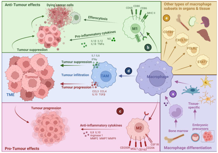
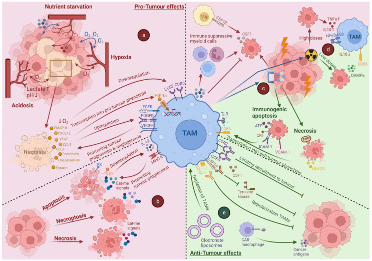
Tumour-associated macrophages (TAMs) are essential players in the tumour microenvironment (TME) and modulate various pro-tumorigenic functions such as immunosuppression, angiogenesis, cancer cell proliferation, invasion and metastasis, along with resistance to anti-cancer therapies. TAMs also mediate important anti-tumour functions and can clear dying cancer cells via efferocytosis. Thus, not surprisingly, TAMs exhibit heterogeneous activities and functional plasticity depending on the type and context of cancer cell death that they are faced with. This ultimately governs both the pro-tumorigenic and anti-tumorigenic activity of TAMs, making the interface between TAMs and dying cancer cells very important for modulating cancer growth and the efficacy of chemo-radiotherapy or immunotherapy. In this review, we discuss the interface of TAMs with cancer cell death from the perspectives of cell death pathways, TME-driven variations, TAM heterogeneity and cell-death-inducing anti-cancer therapies. We believe that a better understanding of how dying cancer cells influence TAMs can lead to improved combinatorial anti-cancer therapies, especially in combination with TAM-targeting immunotherapies.
Keywords: TAM heterogeneity; apoptosis macrophage targeting; cancer therapy; chemotherapy; immunogenic cell death; immunotherapy; macrophages; radiotherapy; tumour microenvironment.
Conflict of interest statement
Abhishek D Garg received consulting/advisory/lecture honoraria from Boehringer Ingelheim (Germany), Miltenyi Biotec (Germany), Novigenix (Switzerland), and IsoPlexis (USA).
Figures
Similar articles
-
Tumour-associated macrophages heterogeneity drives resistance to clinical therapy.Expert Rev Mol Med. 2022 Apr 11;24:e17. doi: 10.1017/erm.2022.8. Expert Rev Mol Med. 2022. PMID: 35400355 Free PMC article. Review.
-
Tumor-associated macrophages: A promising target for a cancer immunotherapeutic strategy.Pharmacol Res. 2020 Nov;161:105111. doi: 10.1016/j.phrs.2020.105111. Epub 2020 Oct 13. Pharmacol Res. 2020. PMID: 33065284 Review.
-
The role of macrophage in regulating tumour microenvironment and the strategies for reprogramming tumour-associated macrophages in antitumour therapy.Eur J Cell Biol. 2021 Mar;100(2):151153. doi: 10.1016/j.ejcb.2021.151153. Epub 2021 Jan 13. Eur J Cell Biol. 2021. PMID: 33476912 Review.
-
Targeting tumor-associated macrophages for cancer immunotherapy.Int Rev Cell Mol Biol. 2022;368:61-108. doi: 10.1016/bs.ircmb.2022.02.002. Epub 2022 Apr 27. Int Rev Cell Mol Biol. 2022. PMID: 35636930
-
Reprogramming tumor-associated macrophages as a unique approach to target tumor immunotherapy.Front Immunol. 2023 Apr 17;14:1166487. doi: 10.3389/fimmu.2023.1166487. eCollection 2023. Front Immunol. 2023. PMID: 37138860 Free PMC article. Review.
Cited by
-
Lymph node and tumor-associated PD-L1+ macrophages antagonize dendritic cell vaccines by suppressing CD8+ T cells.Cell Rep Med. 2024 Jan 16;5(1):101377. doi: 10.1016/j.xcrm.2023.101377. Cell Rep Med. 2024. PMID: 38232703 Free PMC article.
-
The Role of Macrophages in Sarcoma Tumor Microenvironment and Treatment.Cancers (Basel). 2023 Nov 5;15(21):5294. doi: 10.3390/cancers15215294. Cancers (Basel). 2023. PMID: 37958467 Free PMC article. Review.
-
Tp53 determines the spatial dynamics of M1/M2 tumor-associated macrophages and M1-driven tumoricidal effects.Cell Death Dis. 2025 Jan 22;16(1):38. doi: 10.1038/s41419-025-07346-0. Cell Death Dis. 2025. PMID: 39843434 Free PMC article.
-
Trial watch: chemotherapy-induced immunogenic cell death in oncology.Oncoimmunology. 2023 Jun 3;12(1):2219591. doi: 10.1080/2162402X.2023.2219591. eCollection 2023. Oncoimmunology. 2023. PMID: 37284695 Free PMC article. Review.
-
An Epic Advancement in Targeting Macrophages for Cancer Therapy Approach.Curr Drug Deliv. 2025;22(6):732-745. doi: 10.2174/0115672018299798240403062508. Curr Drug Deliv. 2025. PMID: 38591208 Review.
References
-
- Hadadi E., Deschoemaeker S., Venegas G.V., Laoui D. Heterogeneity and function of macrophages in the breast during homeostasis and cancer. Int. Rev. Cell Mol. Biol. 2022;367:149–182. - PubMed
Publication types
MeSH terms
LinkOut - more resources
Full Text Sources
Medical
