. 2022 Dec 2;11(23):3905.
doi: 10.3390/cells11233905.

Genomewide m6A Mapping Uncovers Dynamic Changes in the m6A Epitranscriptome of Cisplatin-Treated Apoptotic HeLa Cells

Affiliations

Genomewide m6A Mapping Uncovers Dynamic Changes in the m6A Epitranscriptome of Cisplatin-Treated Apoptotic HeLa Cells

Azime Akçaöz Alasar et al. Cells. .

Abstract

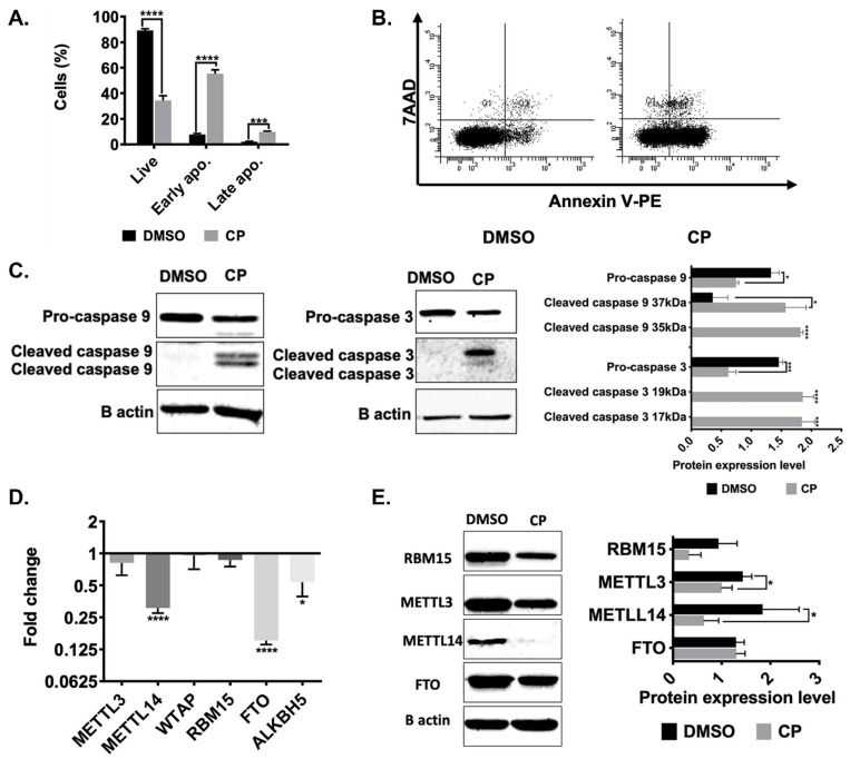
Cisplatin (CP), which is a conventional cancer chemotherapeutic drug, induces apoptosis by modulating a diverse array of gene regulatory mechanisms. However, cisplatin-mediated changes in the m6A methylome are unknown. We employed an m6A miCLIP-seq approach to investigate the effect of m6A methylation marks under cisplatin-mediated apoptotic conditions on HeLa cells. Our high-resolution approach revealed numerous m6A marks on 972 target mRNAs with an enrichment on 132 apoptotic mRNAs. We tracked the fate of differentially methylated candidate mRNAs under METTL3 knockdown and cisplatin treatment conditions. Polysome profile analyses revealed perturbations in the translational efficiency of PMAIP1 and PHLDA1 transcripts. Congruently, PMAIP1 amounts were dependent on METTL3. Additionally, cisplatin-mediated apoptosis was sensitized by METTL3 knockdown. These results suggest that apoptotic pathways are modulated by m6A methylation events and that the METTL3-PMAIP1 axis modulates cisplatin-mediated apoptosis in HeLa cells.

Keywords: HeLa; apoptosis; cisplatin; epitranscriptomics; m6A.


Conflict of interest statement

The authors declare no conflict of interest.

Figures

Figure 1
Figure 1
Expression patterns of m6A enzymes in cisplatin-treated HeLa cells. 1× 10⁶ HeLa cells were treated with 80 μM cisplatin (CP) and 0.1% (v/v) DMSO for 16 h. The cells were stained with Annexin V/7AAD and examined by flow cytometry. Population distributions of DMSO- and CP-treated HeLa cells are depicted (A) Percentage of live (34%), early (55%) and late apoptosis (9.9%) and (B) Dot-blot analysis by flow cytometry after staining with Annexin V-PE and 7AAD. (C) Western blot analysis of caspase- 3 and caspase- 9 in HeLa cells treated with 80 µM CP and 0.1% (v/v) DMSO for 16 h. Equal amounts of total proteins (25 μg/lane) were fractionated through a 10% SDS-PAGE. (D) qPCR analysis of gene expression. Results were normalized against GAPDH. (E) Western blot analysis. Band intensities were normalized against β-actin, used as a loading control. Experiments were conducted in triplicates. *: p ≤ 0.05, ***: p ≤ 0.001, ****: p ≤ 0.0001 by a two-tailed unpaired t-test.
Figure 2
Figure 2
m6A RNA methylome profile of cisplatin-treated HeLa cells. The m6A methylome of DMSO control and CP-treated HeLa cells were obtained by miCLIP-seq as described in Materials and Methods. Distribution of upregulated (A) and downregulated (B) m6A peaks are shown across all transcripts. Pie chart of upregulated (C) and downregulated (D) m6A peaks representing their location on transcripts. Biological replicates: n  =  3 per group. (E) Gene ontology (GO) analysis of differentially m6A-methylated transcripts associated with apoptosis. All biological processes were plotted having a false discovery rate (FDR) <  0.05. (F) m6A methylation profile of PMAIP1.
Figure 3
Figure 3
RNA abundance in cisplatin-treated METTL3 knockdown HeLa cells. (A) Western blot showing METTL3 knockdown in HeLa cells. si-NC was used as negative control. Β-actin was used as loading control. (B) Apoptotic population distribution of si-NC- and si-METTL3-transfected cells followed by DMSO (0.05%)- and CP-treatment (40 µM) for 16 h, respectively. qPCR analyses of total RNAs isolated from DMSO- and CP-treated (C), si-NC and si-METTL3-transfected cells (D), si-METTL3-transfected and DMSO-treated cells (E) and si-METTL3-transfected and CP-treated cells (F). n = 3 p > 0.05, *: p ≤ 0.05, **: p ≤ 0.01 by two-tailed unpaired t-test.
Figure 4
Figure 4
Polysome profiling in METTL3 knockdown HeLa cells. Polysome profiles of cells transfected with si-METTL3 or negative siRNA (si-NC) (A). Total RNAs were phenol-extracted from each fraction collected based on the polysome profile and transcript abundance was measured by qPCR for PHLDA1 (B), PMAIP1 (C), TRAP1 (D) and PIDD1 (E). n = 3. p > 0.05, *: p ≤ 0.05, **: p ≤ 0.01 by two-tailed unpaired t-test.
Figure 5
Figure 5
Polysome profiling in cisplatin-treated METTL3 knockdown HeLa cells. Polysome profiles of cells transfected with si-METTL3 or negative siRNA (si-NC) and treated with 40 µM CP for 16 h (A). Total RNAs were phenol-extracted from each fraction and transcript abundance was examined by qPCR for PHLDA1 (B,C), PMAIP1 (D,E), TRAP1 (F,G) and PIDD1 (H,I) n = 3. *: p ≤ 0.05, **: p ≤ 0.01, ****: p ≤ 0.0001 by two-tailed unpaired t-test.
Figure 5
Figure 5
Polysome profiling in cisplatin-treated METTL3 knockdown HeLa cells. Polysome profiles of cells transfected with si-METTL3 or negative siRNA (si-NC) and treated with 40 µM CP for 16 h (A). Total RNAs were phenol-extracted from each fraction and transcript abundance was examined by qPCR for PHLDA1 (B,C), PMAIP1 (D,E), TRAP1 (F,G) and PIDD1 (H,I) n = 3. *: p ≤ 0.05, **: p ≤ 0.01, ****: p ≤ 0.0001 by two-tailed unpaired t-test.
Figure 6
Figure 6
The METTL3-PMAIP1 axis in METTL3 knockdown and overexpressed HeLa cells. Constitutive PMAIP1 protein expression in HeLa (A,C) and ME-180 (B) cells transfected with si-METTL3 and treated with CP as in Figure 3. Overexpression of METTL3 causes reduction in CP-induced PMAIP1 (D). β-actin was used as loading control. n = 3 *: p ≤ 0.05 ***: p ≤ 0.001, ****: p ≤ 0.0001 by two-tailed unpaired t-test.
Figure 6
Figure 6
The METTL3-PMAIP1 axis in METTL3 knockdown and overexpressed HeLa cells. Constitutive PMAIP1 protein expression in HeLa (A,C) and ME-180 (B) cells transfected with si-METTL3 and treated with CP as in Figure 3. Overexpression of METTL3 causes reduction in CP-induced PMAIP1 (D). β-actin was used as loading control. n = 3 *: p ≤ 0.05 ***: p ≤ 0.001, ****: p ≤ 0.0001 by two-tailed unpaired t-test.
Figure 7
Figure 7
Working model. Under control conditions, PMAIP1 mRNA possesses m6A marks at five different sites. Cisplatin treatment downregulates METTL3 and mediates demethylation of three m6A residues. However, 5′UTR of PMAIP1 is methylated and PMAIP1 translation is enhanced. Translationally enhanced PMAIP1 may then promote apoptosis by inhibiting MCL1 [41], suggesting a novel METTL3–PMAIP1 axis that may modulate apoptosis under cisplatin treatment conditions. * Fold of induction.

Similar articles

Cited by

References

    1. Elmore S. Apoptosis: A review of programmed cell death. Toxicol. Pathol. 2007;35:495–516. doi: 10.1080/01926230701320337. - DOI - PMC - PubMed
    1. Chen P., Li J., Chen Y.C., Qian H., Chen Y.J., Su J.Y., Wu M., Lan T. The functional status of DNA repair pathways determines the sensitization effect to cisplatin in non-small cell lung cancer cells. Cell. Oncol. 2016;39:511–522. doi: 10.1007/s13402-016-0291-7. - DOI - PubMed
    1. Ming X., Groehler I.A., Michaelson-Richie E.D., Villalta P.W., Campbell C., Tretyakova N.Y. Mass Spectrometry Based Proteomics Study of Cisplatin-Induced DNA–Protein Cross-Linking in Human Fibrosarcoma (HT1080) Cells. Chem. Res. Toxicol. 2017;30:980–995. doi: 10.1021/acs.chemrestox.6b00389. - DOI - PMC - PubMed
    1. Wang J., Thomas H.R., Li Z., Yeo N.C., Scott H.E., Dang N., Hossain M.I., Andrabi S.A., Parant J.M. Puma, noxa, p53, and p63 differentially mediate stress pathway induced apoptosis. Cell Death Dis. 2021;12:1–11. doi: 10.1038/s41419-021-03902-6. - DOI - PMC - PubMed
    1. Wei C.-M., Gershowitz A., Moss B. Methylated nucleotides block 5′ terminus of HeLa cell messenger RNA. Cell. 1975;4:379–386. doi: 10.1016/0092-8674(75)90158-0. - DOI - PubMed