Review
. 2022 Nov 23;14(23):5748.
doi: 10.3390/cancers14235748.

The microRNA Lifecycle in Health and Cancer

Affiliations
Review

The microRNA Lifecycle in Health and Cancer

Laura Adriana de Rooij et al. Cancers (Basel). .

Abstract

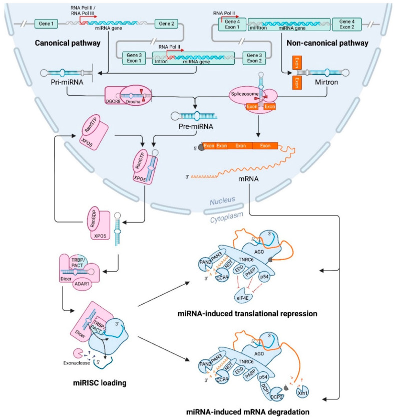
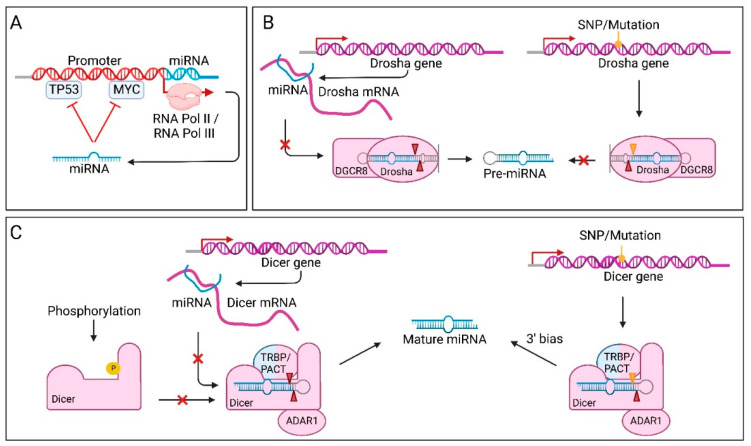
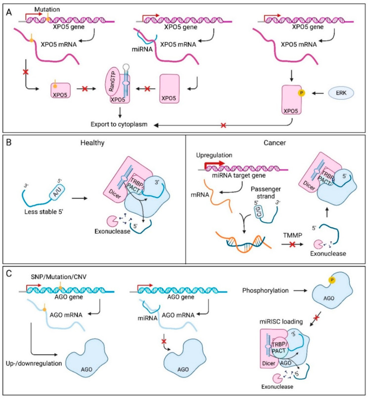
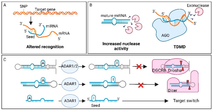
MicroRNAs (miRNAs) are small non-coding RNAs of ~22 nucleotides that regulate gene expression at the post-transcriptional level. They can bind to around 60% of all protein-coding genes with an average of 200 targets per miRNA, indicating their important function within physiological and pathological cellular processes. miRNAs can be quickly produced in high amounts through canonical and non-canonical pathways that involve a multitude of steps and proteins. In cancer, miRNA biogenesis, availability and regulation of target expression can be altered to promote tumour progression. This can be due to genetic causes, such as single nucleotide polymorphisms, epigenetic changes, differences in host gene expression, or chromosomal remodelling. Alternatively, post-transcriptional changes in miRNA stability, and defective or absent components and mediators of the miRNA-induced silencing complex can lead to altered miRNA function. This review provides an overview of the current knowledge on the lifecycle of miRNAs in health and cancer. Understanding miRNA function and regulation is fundamental prior to potential future application of miRNAs as cancer biomarkers.

Keywords: biogenesis; biomarkers; cancer; genetic alterations; microRNA; post-translational regulation.


Conflict of interest statement

The authors declare no conflict of interest.

Figures

Figure 1
Figure 1
General overview of the canonical and non-canonical route from microRNA gene to regulation of target messenger RNA. Following the canonical pathway of miRNA biogenesis, an intergenic (left), or intragenic (middle) miRNA gene is transcribed by RNA polymerase (Pol) II or III, to a primary-miRNA (pri-miRNA) transcript. This stem-loop transcript is cleaved by ribonuclease III enzyme Drosha and its co-factor DiGeorge Syndrome Critical Region 8 (DGCR8) to form a precursor-miRNA (pre-miRNA), which is subsequently exported out of the nucleus by Exportin-5 (XPO5) and its co-factor Ran-GTP. The non-canonical pathway of miRNA biogenesis involves the splicing of a pre-miRNA molecule from an entire intron without the need for Drosha processing, after which it enters the canonical pathway. In the cytoplasm, the pre-miRNA is cleaved by ribonuclease III enzyme Dicer and its co-factors transactivation-response element RNA binding protein (TRBP), protein kinase RNA activator (PACT) and adenosine deaminases acting on RNA 1 (ADAR1) to form a mature miRNA duplex. After strand selection, a single-stranded guide miRNA is incorporated into Argonaute (AGO), which is bound by trinucleotide repeat containing 6 protein (TNRC6) and then the functional miRNA-induced silencing complex (miRISC) is formed. The passenger strand is released and degraded by an exonuclease. After association with multiple co-factors and miRISC effector proteins, the miRISC is able to silence its target. The miRNA is hereby used as a guide, and based on complementarity between miRNA and messenger RNA (mRNA), translation is repressed or the mRNA is degraded. Binding of the miRISC to the target mRNA can induces deadenylation by CCR4-NOT as main effector, which is in some cases followed by decapping by DCP1/DCP2 and degradation by exonuclease XRN1 (Created with BioRender.com).
Figure 2
Figure 2
Genetic factors affecting microRNA expression and function in cancer. (A) Single nucleotide polymorphisms (SNPs) affecting the microRNA (miRNA) gene can alter miRNA production through impeding processing by ribonuclease III enzyme Drosha or Dicer, by alteration in binding and cleavage sites (red triangles). SNPs can also change the seed sequence, leading to a different targetome for the miRNA and thus altered post-translational regulation. (B) In addition to mutations in the miRNA gene itself, the promoter and transcription-enhancing region of miRNA genes can also acquire SNPs. This can impair binding of transcription factors, such as MYC, leading to decreased transcription of the miRNA gene. (C) Epigenetic modifications of the miRNA gene can also affect miRNA expression. Hypermethylation of the miRNA promoter, as well as histone methylation at H3K27, both repress transcription. On the other hand, histone methylation at H3K4 is associated with transcriptional activation of miRNA genes. (D) Some miRNA genes do not have a promoter of their own, which makes them dependent on their host gene. When the host gene promoter is hypermethylated, expression of the miRNA/mirtron is decreased as well. (E) Copy number variations are large chromosomal changes, which include deletions of miRNA genes, or translocations to other positions in the genome, creating gene amplification. When a part of the chromosome is amplified or deleted, the level of miRNA gene expression is decreased or increased, respectively (Created with BioRender.com).
Figure 3
Figure 3
The effect of altered transcription and altered processing through ribonuclease III enzymes Drosha and Dicer malfunctioning. This figure shows how steps of the biogenesis affect production of microRNA (miRNA). (A) Often, miRNAs are transcribed by RNA Polymerase II, which requires transcription factors, such as TP53 and MYC. These transcription factors can be targets of the transcribed miRNA, resulting in a feedback loop. (B) The Drosha RNase III enzyme processes primary miRNAs to precursor miRNAs (pre-miRNA). The protein’s functionality can be altered by many factors, such as single nucleotide polymorphisms (SNPs) and mutations. Drosha messenger RNA can also be the target of specific miRNAs, leading to a negative feedback loop in miRNA maturation. (C) The Dicer RNase III enzyme is able to process pre-miRNA to form a mature miRNA duplex. SNPs and mutations in the Dicer gene can alter its function, leading to altered pre-miRNA processing. Mutations in the RIIID domain of Dicer lead to problems with 5′ strand processing, inducing a 3′ strand bias. Besides inhibition, some SNPs are also capable of increasing Dicer effectivity. Dicer can also be regulated by specific miRNAs, leading to lower miRNA maturation. Phosphorylation of Dicer can induce nuclear translocation, leading to altered miRNA processing in the cytosol (Created with BioRender.com).
Figure 4
Figure 4
Defective microRNA (miRNA) export, miRNA-induced silencing complex (miRISC) loading and Argonaute (AGO) malfunction, leading to altered miRNA functionality in cancer. (A) Exportin-5 (XPO5) exports precursor miRNAs (pre-miRNAs) from the nucleus to the cytoplasm. When the XPO5 gene is mutated, pre-miRNA is retained in the nucleus, resulting in lowered mature miRNA levels. XPO5 functionality can also be altered through post-translational regulation by specific miRNAs. Moreover, phosphorylation by extracellular signal-regulated kinase (ERK) inhibits the shuttling of XPO5 to the cytosol, leading to decreased levels of pre-miRNA in the cytoplasm. (B) Loading of one of the miRNA strands into the miRNA-induced silencing complex (miRISC) determines targetome that will be regulated. Strand selection is determined by least 5′ end thermodynamics and preferably adenine or uracil nucleotides. The strand that meets these requirements the most, is implemented, whilst the other is degraded. However, when the interaction of the discarded strand with its own target mRNA is strong, this interaction prevents miRNA degradation, leading to arm switching. This mechanism is called target-mediated miRNA protection (TMMP). (C) The assembly of the miRISC can also be regulated through differences in AGO proteins. Transcription of the AGO genes can be altered through single nucleotide polymorphisms, mutations or copy number variations (CNVs) in cancer, leading to de- or increased miRNA functionality. AGO can also be targeted by specific miRNAs, creating a negative feedback loop. Moreover, the AGO proteins can be phosphorylated, leading to impaired miRISC loading (Created with BioRender.com).
Figure 5
Figure 5
microRNA dysregulation in cancer through target gene modifications, and factors affecting microRNA degradation and microRNA editing. (A) A mutation in the microRNA (miRNA) recognition site of a target gene can result in multiple changes; the target can become undetectable for the miRNA, the mutation might change the degree of repression or make a target recognizable for miRNA. (B) miRNA stability in the cell is important for regulation of miRNA targets, and stability is lowered by increased nuclease activity or target-directed miRNA degradation (TDMD). The latter process, TDMD, revolves around complementarity between target and miRNA. When a miRNA binds to its target with high 3′ end complementarity, the conformation of Argonaute (AGO) may change and the miRNA may become susceptible to degradation by exonucleases. (C) RNA editing, such as adenosine (A) to inosine (I) editing by adenosine deaminases acting on RNA 1/2 (ADAR1/2), can change the sequence of the RNA, with diverse consequences. miRNA editing can alter primary-miRNA and pre-miRNA in such a way, that they become unavailable for processing by ribonuclease III enzymes Drosha and Dicer, respectively. Additionally, the seed can also be edited, which allows for a shift in targetome, or altered recognition of existing targets (Created with BioRender.com).

Similar articles

Cited by

References

    1. Wightman B., Ha I., Ruvkun G. Posttranscriptional Regulation of the Heterochronic Gene Lin-14 by Lin-4 Mediates Temporal Pattern Formation in C. Elegans. Cell. 1993;75:855–862. doi: 10.1016/0092-8674(93)90530-4. - DOI - PubMed
    1. Lee R.C., Ambros V. An Extensive Class of Small RNAs in Caenorhabditis Elegans. Science. 2001;294:862–864. doi: 10.1126/science.1065329. - DOI - PubMed
    1. Caudy A.A., Myers M., Hannon G.J., Hammond S.M. Fragile X-Related Protein and VIG Associate with the RNA Interference Machinery. Genes Dev. 2002;16:2491–2496. doi: 10.1101/gad.1025202. - DOI - PMC - PubMed
    1. Lewis B.P., Shih I., Jones-Rhoades M.W., Bartel D.P., Burge C.B. Prediction of Mammalian MicroRNA Targets. Cell. 2003;115:787–798. doi: 10.1016/S0092-8674(03)01018-3. - DOI - PubMed
    1. Friedman R.C., Farh K.K.-H., Burge C.B., Bartel D.P. Most Mammalian MRNAs Are Conserved Targets of MicroRNAs. Genome Res. 2009;19:92–105. doi: 10.1101/gr.082701.108. - DOI - PMC - PubMed

LinkOut - more resources