

MicroRNA-Gene Interactions Impacted by Toxic Metal(oid)s during EMT and Carcinogenesis
- PMID: 36497298
- PMCID: PMC9741118
- DOI: 10.3390/cancers14235818
MicroRNA-Gene Interactions Impacted by Toxic Metal(oid)s during EMT and Carcinogenesis
Abstract
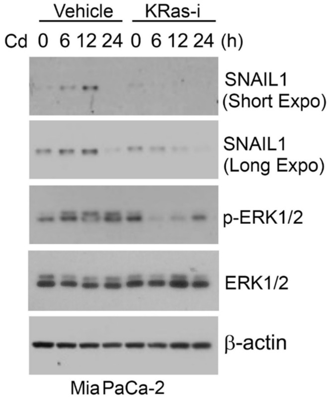
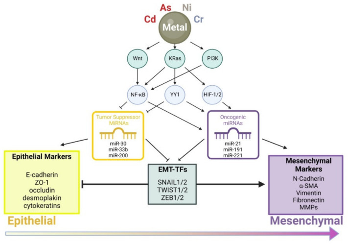
Chronic environmental exposure to toxic metal(loid)s significantly contributes to human cancer development and progression. It is estimated that approximately 90% of cancer deaths are a result of metastasis of malignant cells, which is initiated by epithelial-mesenchymal transition (EMT) during early carcinogenesis. EMT is regulated by many families of genes and microRNAs (miRNAs) that control signaling pathways for cell survival, death, and/or differentiation. Recent mechanistic studies have shown that toxic metal(loid)s alter the expression of miRNAs responsible for regulating the expression of genes involved in EMT. Altered miRNA expressions have the potential to be biomarkers for predicting survival and responses to treatment in cancers. Significantly, miRNAs can be developed as therapeutic targets for cancer patients in the clinic. In this mini review, we summarize key findings from recent studies that highlight chemical-miRNA-gene interactions leading to the perturbation of EMT after exposure to toxic metal(loid)s including arsenic, cadmium, nickel, and chromium.
Keywords: EMT; arsenic; cadmium; carcinogenesis; chromium; metals; microRNAs; nickel.
Conflict of interest statement
The authors declare no conflict of interest.
Figures
References
Publication types
Grants and funding
LinkOut - more resources
Full Text Sources
