

Diagnostics Using Non-Invasive Technologies in Dermatological Oncology
- PMID: 36497368
- PMCID: PMC9738560
- DOI: 10.3390/cancers14235886
Diagnostics Using Non-Invasive Technologies in Dermatological Oncology
Abstract
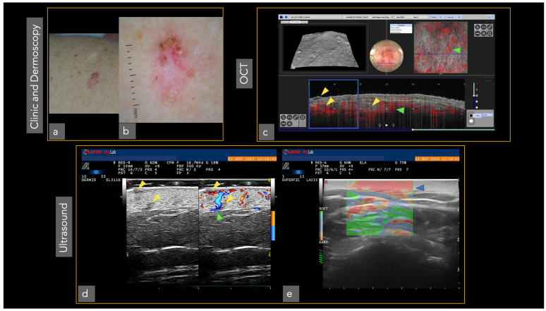
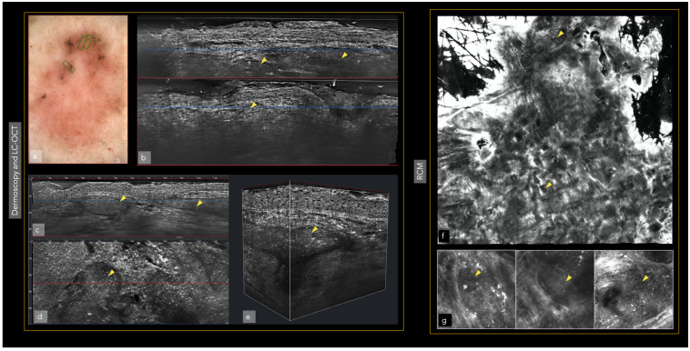
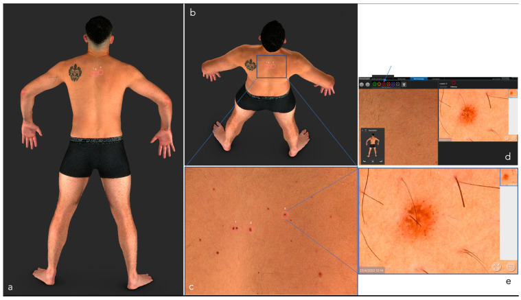
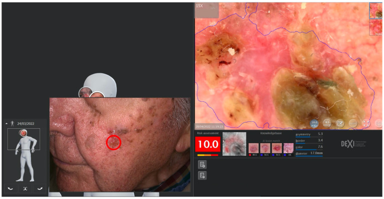
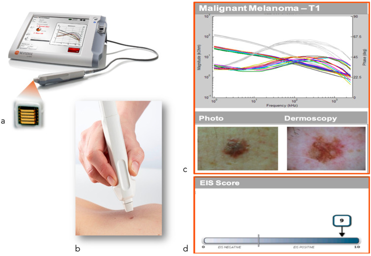
The growing incidence of skin cancer, with its associated mortality and morbidity, has in recent years led to the developing of new non-invasive technologies, which allow an earlier and more accurate diagnosis. Some of these, such as digital photography, 2D and 3D total-body photography and dermoscopy are now widely used and others, such as reflectance confocal microscopy and optical coherence tomography, are limited to a few academic and referral skin cancer centers because of their cost or the long training period required. Health care professionals involved in the treatment of patients with skin cancer need to know the implications and benefits of new non-invasive technologies for dermatological oncology. In this article we review the characteristics and usability of the main diagnostic imaging methods available today.
Keywords: LC-OCT; OCT; RCM; artificial intelligence; diagnostics; high frequency ultrasound; imaging; imaging techniques; skin cancer; total body photography (TBP).
Conflict of interest statement
The authors declare no conflict of interest.
Figures
Similar articles
-
[Modern diagnostic procedures in dermatological oncology].Pathologe. 2015 Feb;36(1):11-5. doi: 10.1007/s00292-014-2062-4. Pathologe. 2015. PMID: 25630484 Review. German.
-
Role of In Vivo Reflectance Confocal Microscopy in the Analysis of Melanocytic Lesions.Acta Dermatovenerol Croat. 2018 Apr;26(1):64-67. Acta Dermatovenerol Croat. 2018. PMID: 29782304 Review.
-
Practice Gaps in Dermatology: Melanocytic Lesions and Melanoma.Dermatol Clin. 2016 Jul;34(3):353-62. doi: 10.1016/j.det.2016.03.003. Dermatol Clin. 2016. PMID: 27363893 Review.
-
Features of tattoo-associated cutaneous lymphoid hyperplasia on reflectance confocal microscopy and line-field confocal optical coherence tomography.Australas J Dermatol. 2024 May;65(3):e50-e55. doi: 10.1111/ajd.14246. Epub 2024 Mar 4. Australas J Dermatol. 2024. PMID: 38439201
-
Update on non-invasive imaging techniques in early diagnosis of non-melanoma skin cancer.G Ital Dermatol Venereol. 2015 Aug;150(4):393-405. Epub 2015 Jul 16. G Ital Dermatol Venereol. 2015. PMID: 26184797 Review.
Cited by
-
Multispectral Imaging for Skin Diseases Assessment-State of the Art and Perspectives.Sensors (Basel). 2023 Apr 11;23(8):3888. doi: 10.3390/s23083888. Sensors (Basel). 2023. PMID: 37112229 Free PMC article. Review.
-
Dermoscopy and reflectance confocal microscopy of solitary flat pink lesions: A new combined score to diagnose amelanotic melanoma.J Eur Acad Dermatol Venereol. 2025 Jan;39(1):109-116. doi: 10.1111/jdv.19991. Epub 2024 Apr 4. J Eur Acad Dermatol Venereol. 2025. PMID: 38572809 Free PMC article.
-
Editorial: Methods in skin cancer.Front Oncol. 2023 Mar 3;13:1150450. doi: 10.3389/fonc.2023.1150450. eCollection 2023. Front Oncol. 2023. PMID: 36937449 Free PMC article. No abstract available.
-
Interobserver agreement on line-field confocal optical coherence tomography image markers in keratinocyte carcinomas and precursor lesions.Arch Dermatol Res. 2024 Sep 6;316(8):608. doi: 10.1007/s00403-024-03344-y. Arch Dermatol Res. 2024. PMID: 39240381 Free PMC article.
-
Histology and line-field confocal optical coherence tomography in granuloma faciale: A single case study.J Dtsch Dermatol Ges. 2024 Dec;22(12):1676-1678. doi: 10.1111/ddg.15569. Epub 2024 Oct 9. J Dtsch Dermatol Ges. 2024. PMID: 39382211 Free PMC article. No abstract available.
References
-
- Leiter U., Eigentler T., Garbe C. Epidemiology of skin cancer. Adv. Exp. Med. Biol. 2014;810:120–140. - PubMed
-
- Garbe C., Amaral T., Peris K., Hauschild A., Arenberger P., Basset-Seguin N., Bastholt L., Bataille V., Del Marmol V., Dréno B., et al. European Dermatology Forum (EDF), the European Association of Dermato-Oncology (EADO), and the European Organization for Research and Treatment of Cancer (EORTC). European consensus-based interdisciplinary guideline for melanoma. Part 1: Diagnostics: Update 2022. Eur. J. Cancer. 2022;170:236–255. - PubMed
Publication types
Grants and funding
- PI15/00716/Fondo de Investigaciones Sanitarias (Spain),
- PI15/00956/CIBER de Enfermedades Raras of the Instituto de Salud Carlos III, Spain
- PI18/00419/ISCIII-Subdireccion General de Evaluacion and European Regional Development Fund (ERDF), "a way to make Europe"
- AGAUR 2017_SGR_1134/CERCA Programme by Generalitat de Catalunya, Spain;
- LSHC-CT-2006-018702 (GenoMEL)/European Commission under the 6th Framework Programme
- HORIZON2020 Framework Programme, iTobos and Qualitop/European Commission under the 7th Framework Programme, Diagnoptics, and the European Commision
- (CA83115)/The National Cancer Institute (NCI) of the US National Institute of Health (NIH)
- 201331-30/Fundació La Marató de TV3
- GCB15152978SOEN,/"Fundación Científica de la Asociación Española Contra el Cáncer"
LinkOut - more resources
Full Text Sources
Medical
