. 2022 Nov 30;14(23):5944.
doi: 10.3390/cancers14235944.

A Signaling Crosstalk Links SNAIL to the 37/67 kDa Laminin-1 Receptor Ribosomal Protein SA and Regulates the Acquisition of a Cancer Stem Cell Molecular Signature in U87 Glioblastoma Neurospheres

Affiliations

A Signaling Crosstalk Links SNAIL to the 37/67 kDa Laminin-1 Receptor Ribosomal Protein SA and Regulates the Acquisition of a Cancer Stem Cell Molecular Signature in U87 Glioblastoma Neurospheres

Loraine Gresseau et al. Cancers (Basel). .

Erratum in

Abstract

Background: Three-dimensional in vitro neurospheres cultures recapitulate stemness features associated with poor clinical outcome in glioblastoma patients. They are commonly used to address brain cancer stem cell (CSC) signal transducing biology that regulates spheroids formation and stemness phenotype, and to assess the in vitro pharmacological impact of chemotherapeutic drugs. Objective: Here, we addressed the role of a new signaling axis involved in the regulation of in vitro spheroids formation and assessed the chemopreventive ability of diet-derived epigallocatechin gallate (EGCG) to impact the processes that govern the acquisition of spheroids CSC stemness traits. Methods: Neurospheres were generated from adherent human U87 glioblastoma cancer cell cultures under conditions that recapitulate stemness features. Total RNA and protein lysates were isolated for gene expression by RT-qPCR and protein expression by immunoblot. Transcriptomic analysis was performed through RNA-Seq. Results: Compared to their parental adherent cells, tumorspheres expressed increased levels of the CSC markers NANOG, SOX2, PROM1 (CD133), as well as of the epithelial-to-mesenchymal transition (EMT) markers Fibronectin, SNAI1, and 37/67 kDa laminin-1 receptor ribosomal protein SA (RPSA). Increased PROM1, SOX2, Fibronectin, and RPSA transcripts level were also observed in clinical grade IV glioblastoma tissues compared to normal tissue. EGCG treatment reduced dose-dependently tumorspheres size and inhibited the transcriptional regulation of those genes. An apoptotic signature was also found in spheroids with increased signal transducing events involving GSK3α/β, RSK, and CREB. These were repressed upon RPSA gene silencing and partially by SNAI1 silencing. Conclusion: This work highlights a signaling axis linking RPSA upstream of SNAIL in neurospheres genesis and supports the chemopreventive impact that diet-derived EGCG may exert on the acquisition of CSC traits.

Keywords: EGCG; EMT; RPSA; SNAIL; cancer stem cells; glioblastoma; laminin receptor; spheroids.


Conflict of interest statement

The authors declare no conflict of interest.

Figures

Figure 1
Figure 1
EGCG alters neurospheres size and reverses the acquisition of a cancer-stem cell phenotype. (A) U87 glioblastoma monolayers were cultured with the Tumorsphere Medium Xf with SupplementMix for the indicated times in the absence or presence of increasing EGCG concentrations and spheroids. Representative phase contrast pictures were taken, and (B) spheroid size was quantified as described in the Methods section (**** = 0.0001 ≥ p; ns = non significant) (C) Total RNA was extracted from adherent or neurospheres generated upon 72 h in the presence of increasing EGCG concentrations. RT-qPCR analysis was used to assess the expression of CSC markers PROM1, SOX2, and NANOG. Probability values of 0.05 were judged significant and indicated as (*). (D) Cell lysates were harvested from adherent, or spheroids obtained at 72 h. Western blot analysis of protein expression was performed as described in the Methods section of the described markers. Representative blots for each marker are shown from three independent experiments. (E) In silico analysis of PROM1, SOX2, and NANOG transcript levels was performed on clinical samples from GBM, and low-grade glioma (LGG) and compared to healthy tissue as described in the Methods section. Probability values of 0.05 were judged significant and indicated as (*).
Figure 2
Figure 2
Transcriptomic analysis reveals the reversion of epithelial to mesenchymal transition in profit to an apoptotic program upon EGCG treatment of glioblastoma spheroids. (A) Total RNA was extracted from control- and EGCG- treated U87 spheroids from three independent experiments and subjected to RNA-Sequencing. Unsupervised hierarchical clustering of all the genes differentially expressed in pairwise comparison was tested with adjusted p value ≤ 0.05 and log2 fold change ≥1.0. (B) Dot plot showing changes in the normalized enrichment score (NES) for the most enriched pathways in EGCG-treated U87 spheroids compared to untreated control cells. FDR p value ≤ 0.05. The top up- and down-regulated gene signatures of Hallmark, Canonical pathways, and Gene Ontology. (C) Heatmap representation of the relative expression of known phenotypic markers for each indicated cellular process.
Figure 3
Figure 3
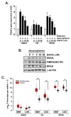
EGCG alters neurospheres’ epithelial-to-mesenchymal transition phenotype. (A) U87 glioblastoma monolayers were cultured with the Tumorsphere Medium Xf with SupplementMix to generate spheroids for 72 h in the absence or presence of increasing EGCG concentrations. Total RNA was extracted from adherent or neurospheres, and RT-qPCR analysis was used to assess the expression of EMT markers SNAIL, Fibronectin (FN), and RPSA (67 kDa Laminin Receptor). (B) Cell lysates were harvested from adherent, or spheroids obtained at 72 h. Western blot analysis of protein expression was performed as described in the Methods section of the described markers. Representative blots for each marker are shown from three independent experiments. (C) In silico analysis of SNAI1, FN, and RPSA transcript levels was performed on clinical samples from GBM, and low-grade glioma (LGG) and compared to healthy tissue as described in the Methods section. Probability values of 0.05 were judged significant and indicated in the figure as (*).
Figure 4
Figure 4
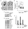
Repression of RPSA alters spheroids formation and prevents the acquisition of a cancer stem cell phenotype. (A) U87 glioblastoma monolayers were transiently transfected with a scrambled sequence (siScrambled) or with a siRNA directed against RPSA (siRPSA). Next, the cells were cultured with the Tumorsphere Medium Xf with Supplement Mix for 72 h in the absence or presence of 30 μM EGCG. Representative phase contrast pictures of the spheroids formed were taken, and (B) spheroid size was quantified as described in the Methods section. (*** = 0.001 ≥ p; **** = 0.0001 ≥ p; ns = non significant). Cell lysates were harvested from adherent, or spheroids obtained at 72 h and upon transient siScrambled or siRPSA transfection. Western blot analysis of (C) EMT markers RPSA, SNAIL, and β-Actin, as well as the indicated (D) apoptosis and transducing intermediates expression was performed as described in the Methods section. Representative blots for each marker are shown from three independent experiments. (E) Total RNA was extracted from adherent or neurospheres and RT-qPCR analysis was used to assess the expression of CSC markers (PROM1, SOX2, and NANOG), and EMT marker SNAI1. Probability values of 0.05 were judged significant and indicated as (*).
Figure 5
Figure 5
Repression of SNAIL alters spheroids formation and prevents the acquisition of a cancer stem cell phenotype but does not affect the acquisition of the apoptotic signature. (A) U87 glioblastoma monolayers were transiently transfected with a scrambled sequence (siScrambled) or with a siRNA directed against SNAIL (siSNAIL). Next, the cells were cultured with the Tumorsphere Medium Xf with SupplementMix for 72 h in the absence or presence of 30 μM EGCG. Representative phase contrast pictures of the spheroids formed were taken, and (B) spheroid size was quantified as described in the Methods section. (*** = 0.001 ≥ p; **** = 0.0001 ≥ p; ns = non significant). Cell lysates were harvested from adherent, or spheroids obtained at 72 h and upon transient siScrambled or siSNAIL transfection. Western blot analysis of (C) EMT markers RPSA, SNAIL, and β-Actin, as well as the indicated (D) apoptosis and transducing intermediates expression was performed as described in the Methods section. Representative blots for each marker are shown from three independent experiments. (E) Total RNA was extracted from adherent or neurospheres, and RT-qPCR analysis was used to assess the expression of CSC markers (PROM1, SOX2, and NANOG). Probability values of 0.05 were judged significant and indicated as (*).
Figure 6
Figure 6
Schematic representation summarizing the crosstalk linking SNAIL to RPSA and the action of EGCG on the neurosphere molecular signature. U87 glioblastoma monolayers (2D cell cultures) can be cultured into 3D neurospheres to recapitulate the CSC invasive and chemo-resistant phenotypes through, in part, the induction of epithelial-to-mesenchymal (EMT) process and adaptative response to low O2 levels (hypoxia). Neurospheres express increased RPSA levels, contributing to both SNAIL-dependent and SNAIL-independent events. EGCG (red arrow) was demonstrated to target SNAIL-dependent signaling by reducing EMT biomarkers, and RSK phosphorylation, to impede spheroids growth and CSC phenotype. A potential adaptative apoptotic phenotype also involves SNAIL-independent signaling where RPSA regulates the phosphorylation status of MSK/CREB/GSK3α/β during spheroids formation. Altogether, current EGCG pharmacological intervention against acquiring of a CSC phenotype supports its chemopreventive properties.

References

    1. Tamimi A.F., Juweid M. Epidemiology and Outcome of Glioblastoma. In: De Vleeschouwer S., editor. Glioblastoma. Codon Publications; Brisbane, Australia: 2017. Chapter 8. - PubMed
    1. Cruz J.V.R., Batista C., Afonso B.H., Alexandre-Moreira M.S., Dubois L.G., Pontes B., Moura Neto V., Mendes F.A. Obstacles to glioblastoma treatment two decades after temozolomide. Cancers. 2022;14:3203. doi: 10.3390/cancers14133203. - DOI - PMC - PubMed
    1. Arvanitis C.D., Ferraro G.B., Jain R.K. The blood-brain barrier and blood-tumour barrier in brain tumours and metastases. Nat. Rev. Cancer. 2020;20:26–41. doi: 10.1038/s41568-019-0205-x. - DOI - PMC - PubMed
    1. Alves A.L.V., Gomes I.N.F., Carloni A.C., Rosa M.N., da Silva L.S., Evangelista A.F., Reis R.M., Silva V.A.O. Role of glioblastoma stem cells in cancer therapeutic resistance: A perspective on antineoplastic agents from natural sources and chemical derivatives. Stem Cell Res. Ther. 2021;12:206. doi: 10.1186/s13287-021-02231-x. - DOI - PMC - PubMed
    1. Sun H.R., Wang S., Yan S.C., Zhang Y., Nelson P.J., Jia H.L., Qin L.X., Dong Q.Z. Therapeutic strategies targeting cancer stem cells and their microenvironment. Front. Oncol. 2019;9:1104. doi: 10.3389/fonc.2019.01104. - DOI - PMC - PubMed