. 2022 Nov 22;11(23):6895.
doi: 10.3390/jcm11236895.

Schober Test and Its Modifications Revisited-What Are We Actually Measuring? Computerized Tomography-Based Analysis

Affiliations

Schober Test and Its Modifications Revisited-What Are We Actually Measuring? Computerized Tomography-Based Analysis

Oded Hershkovich et al. J Clin Med. .

Abstract

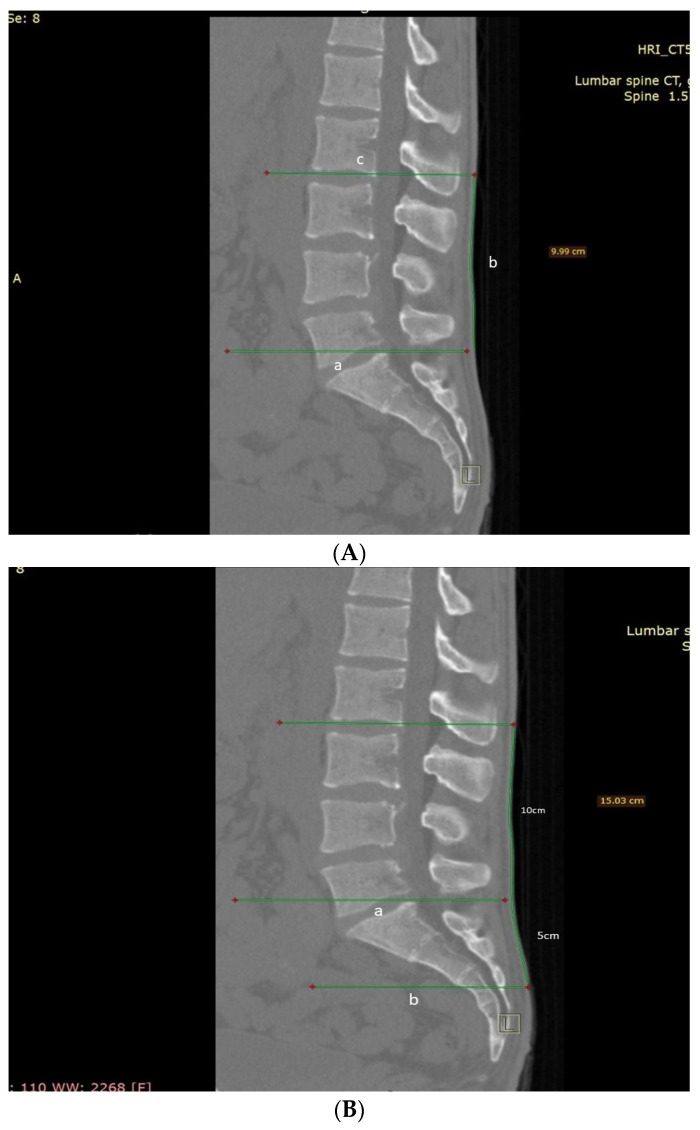
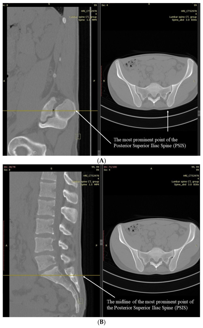
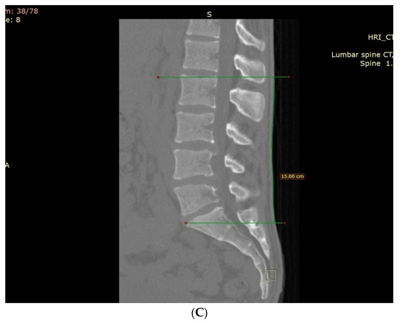
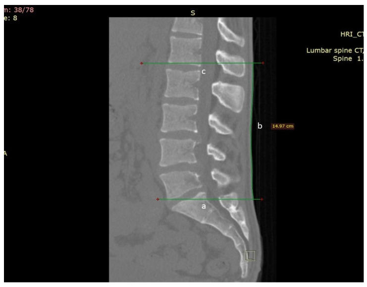
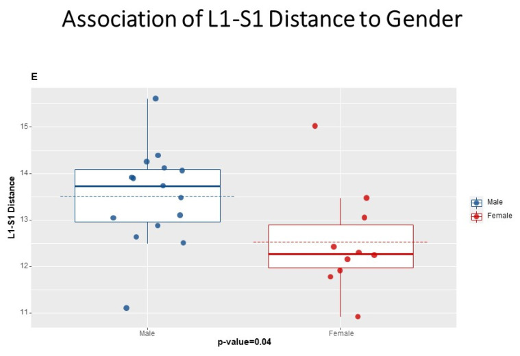
Objective: Examine Schober test's (ST), Modified ST (MST), and Modified-Modified ST (MMST) surface markers' accuracy in spanning lumbar L1-S1 motion segments and repeatability related to actual patient anatomy as measured on sagittal CT scans.

Methods: The study included 25 patients of varying heights, weights, and gender without prior spinal surgery or deformity. Researchers assessed patients' CT scans for ST, MST, and MMST skin levels of the measured cephalic and caudal endpoints.

Results: The original ST failed to include at least one lumbar motion segment in all patients, omitting the L1-L2 motion segment in 17 patients and the L2-L3 in another eight. The additional cephalic length of the MST did not improve the inclusion of the actual L1-S1 components. The MMST measured 19 'patients' entire L1-S1 motion segments, reaching a 76% accuracy rate. WMST, measuring 16 cm (instead of MMST's 15 cm), improved the measurement significantly, measuring the L1-S1 motion segments in all cases (with 100% accuracy).

Conclusion: ST and its modifications fail to span the L1-S1 motion segments and are thus prone to underestimating lumbar spine motion. This study shows that the WMST is much more accurate than previous modifications and is a better tool for evaluating lumbar spine motion.

Keywords: Schober test; lumbar; modified; range of motion.


Conflict of interest statement

All authors have completed the ICMJE uniform disclosure form at www.icmje.org/coi_disclosure.pdf (accessed on 1 November 2022) (available on request from the corresponding author) and declare no conflict of interest.

Figures

Figure 1
Figure 1
(A) Schober Test CT measurement. A parallel line is drawn from the posterosuperior corner of the S1 vertebra to the skin (marked as a), which is followed by a 10 cm line drawn over the skin to reach the cephalad endpoint imitating the original Schober test (marked as b). A parallel line was drawn from this point to the skeletal spine level, transecting a vertebra (upper third, middle third, or lower third) or a disc space (marked as c). Lumbar vertebrae were numbered as (L)1 to (L)5, with the upper third as 0.3, middle third as 0.6, and lower third as 0.9. A line transecting the middle third of L2 was therefore designated as 2.6. Sacral vertebras were numbered from 6 to 10 similarly. (B) Modified Schober Test CT measurement. It was measured similarly to the Schober test by marking a skin point 5 cm caudal and 10 cm cephalad to the lumbosacral junction (a parallel line drawn from the posterosuperior corner of the S1 vertebra to the skin) (Marked as a). Spinal endpoints were measured; the cephalad point was measured as in the Schober test. The caudal end was measured as the parallel line from the caudal 5 cm skin point to the skeletal spine level (upper third, middle third, or lower third of a vertebra or the disc space) (Marked as b).
Figure 2
Figure 2
MMST Measurement. (A) MMST PSIS LANDMARK on CT axial–sagittal views. (B) MMST PSIS Qaudal starting point. MMST was measured by marking the most prominent point of the Posterior Superior Iliac Spine (PSIS), as measured by CT (Marked arrows). On that level, scrolling back to a midline sagittal plane, a parallel line was drawn to the skin surface as described previously. (C) MMST complete CT measurement. From that point, a 15 cm skin surface line was drawn cranially. The caudal and cephalad spinal levels were measured as described before.
Figure 2
Figure 2
MMST Measurement. (A) MMST PSIS LANDMARK on CT axial–sagittal views. (B) MMST PSIS Qaudal starting point. MMST was measured by marking the most prominent point of the Posterior Superior Iliac Spine (PSIS), as measured by CT (Marked arrows). On that level, scrolling back to a midline sagittal plane, a parallel line was drawn to the skin surface as described previously. (C) MMST complete CT measurement. From that point, a 15 cm skin surface line was drawn cranially. The caudal and cephalad spinal levels were measured as described before.
Figure 3
Figure 3
Actual lumbar (L1-S1) motion segments length measurement on CT. A line was stretched from the mid-endplate of S1 to the skin surface (marked as a), and a parallel line was extended from the mid-endplate of L1 to the skin surface (marked as c). The distance between both lines was measured on the skin surface (marked as b).
Figure 4
Figure 4
Association of L1-S1 Distance to Gender.

References

    1. Lotan R., Smorgick Y., Anekstein Y., Rudik O., Prosso I., Hershkovich O. Kyphoplasty for Elderly Patients with Vertebral Compression Fractures—Do We Save Lives? Mortality Rates Analysis Comparison in a Long-Term Follow-Up Cohort. Glob. Spine J. 2022;12:1443–1448. doi: 10.1177/2192568220982282. - DOI - PMC - PubMed
    1. Scura D., Piazza V.M. Spine Secrets Plus. Elsevier; Amsterdam, The Netherlands: 2021. Disability Evaluation.
    1. Jassal N.S. Treatment of Chronic Pain Conditions. Springer; Berlin/Heidelberg, Germany: 2017. Lumbar Spine History and Physical; pp. 33–35.
    1. Doğan M., Koçak M., Kılınç Ö.O., Ayvat F., Sütçü G., Ayvat E., Kılınç M., Ünver Ö., Yıldırım S.A. Functional range of motion in the upper extremity and trunk joints: Nine functional everyday tasks with inertial sensors. Gait Posture. 2019;70:141–147. doi: 10.1016/j.gaitpost.2019.02.024. - DOI - PubMed
    1. Jette A.M., Smith K., Haley S.M., Davis K.D. Physical therapy episodes of care for patients with low back pain. Phys. Ther. 1994;74:101–110. doi: 10.1093/ptj/74.2.101. - DOI - PubMed

LinkOut - more resources