Review
. 2022 Dec 1;11(23):7156.
doi: 10.3390/jcm11237156.

Local Resection in Choroidal Melanoma: A Review

Affiliations
Review

Local Resection in Choroidal Melanoma: A Review

Josep Maria Caminal et al. J Clin Med. .

Abstract

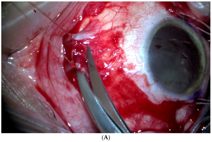
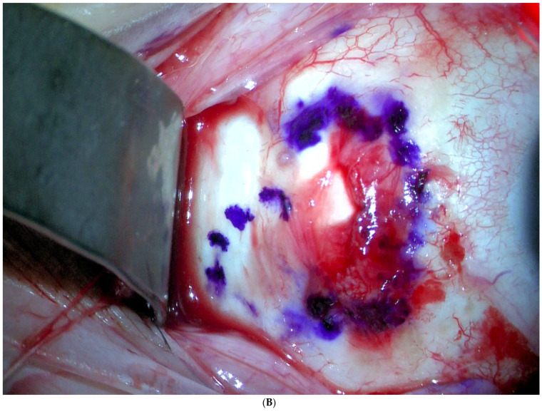
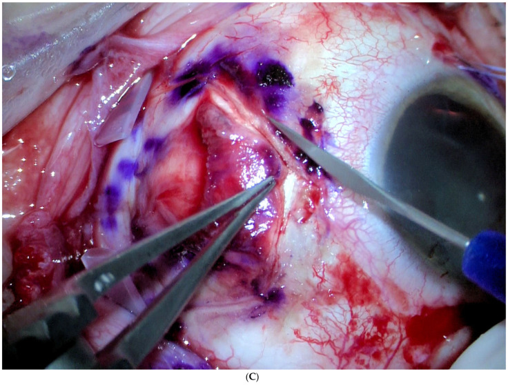
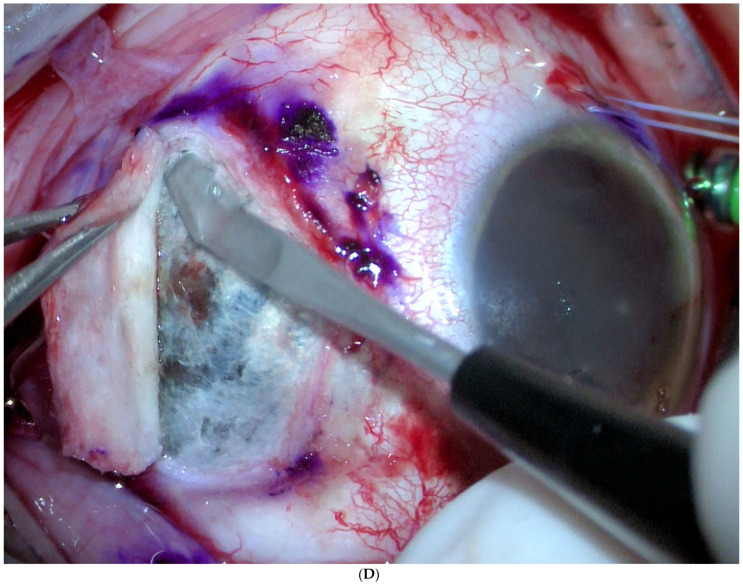
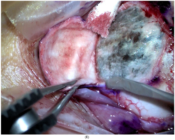
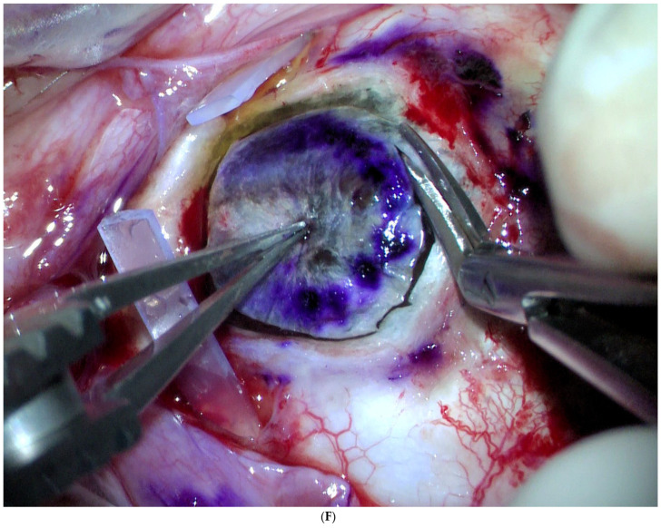
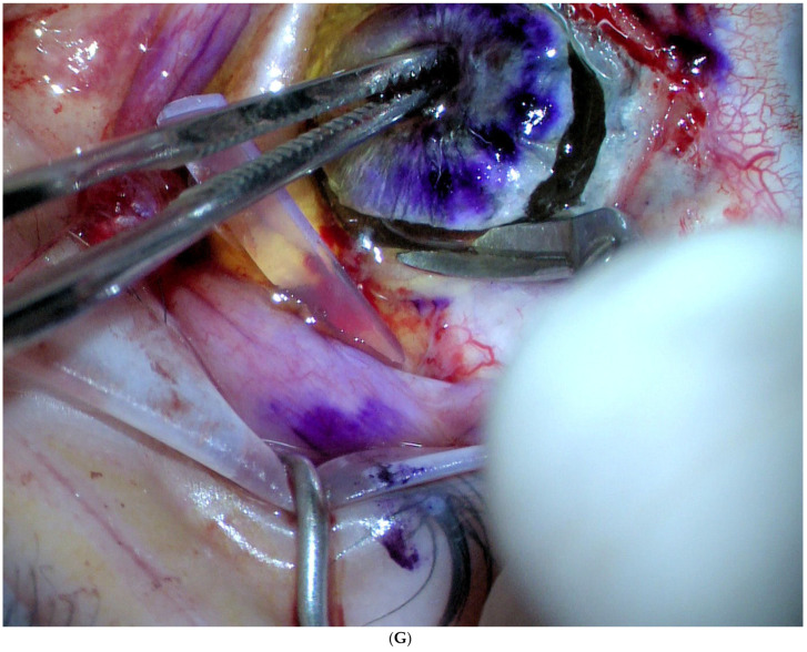
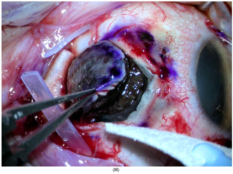
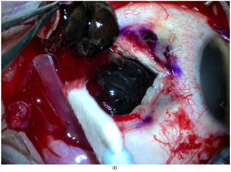
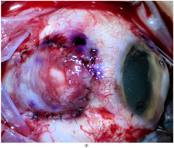
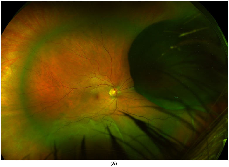
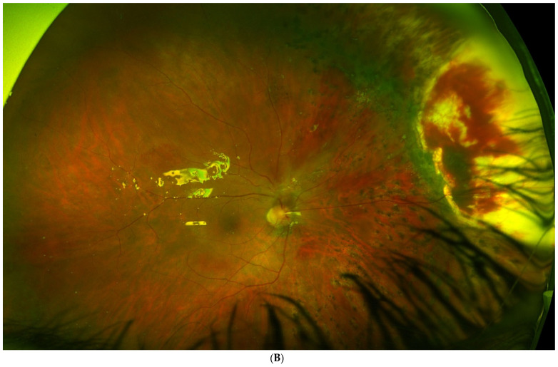
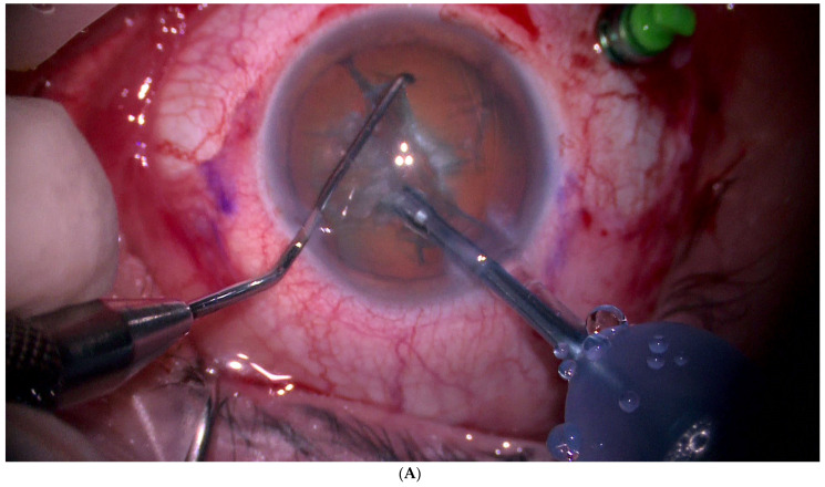
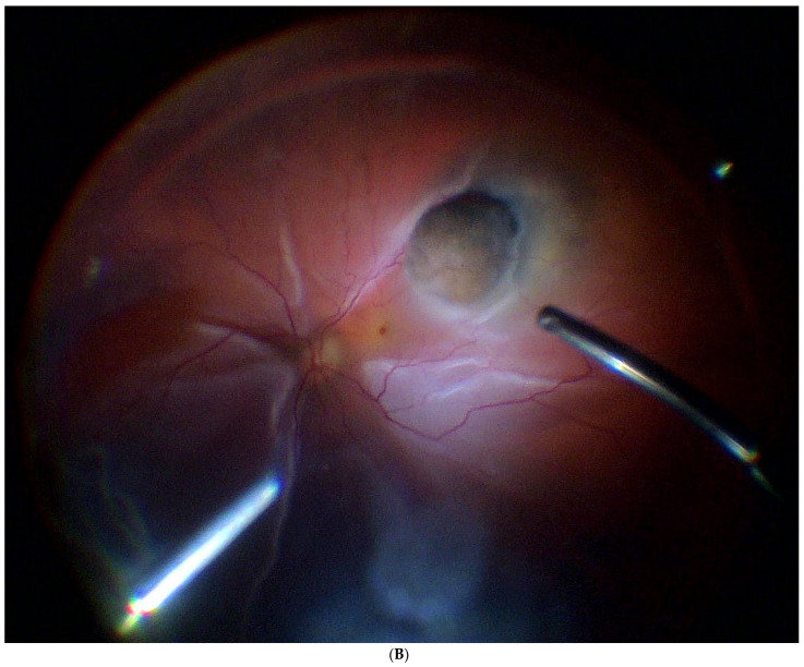
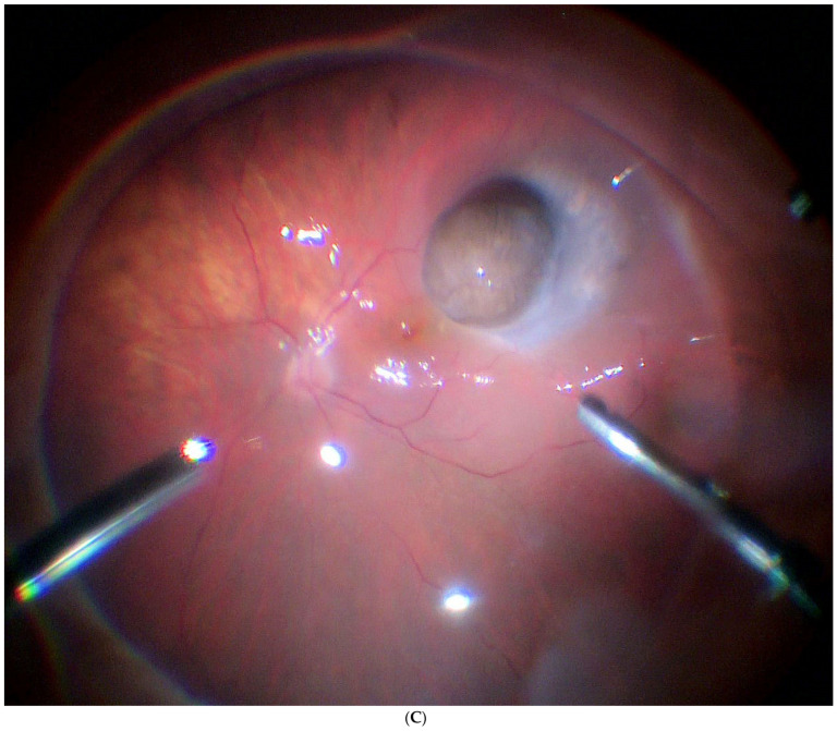
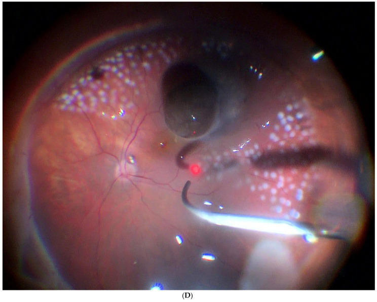
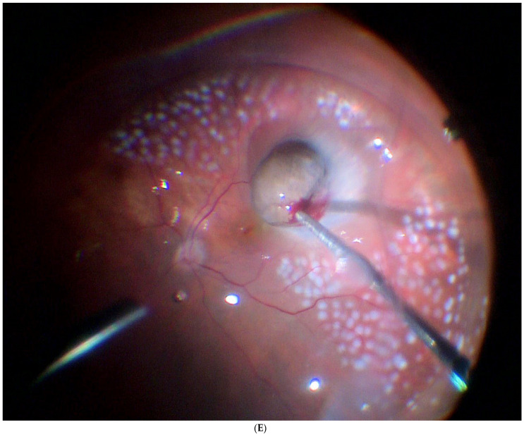
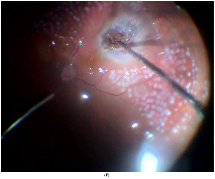
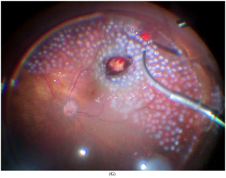
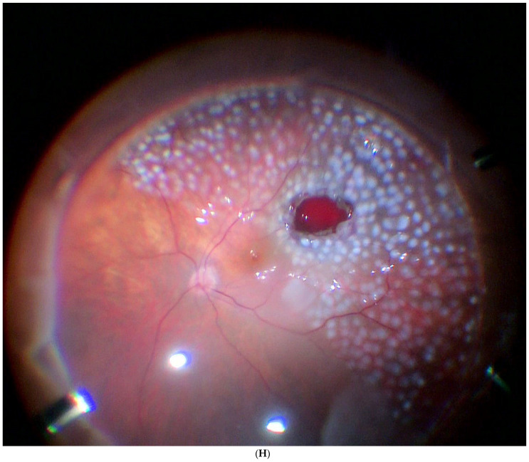
Surgical resection is widely used to treat small tumours located in the iris and the ciliary body, due to the accessibility of these sites. By contrast, surgical removal of choroidal tumours is substantially more challenging, which is why this procedure is performed only at specialised centres. In the present article, we review the literature on surgical resection of choroidal tumours, which can be performed as endoresection (ab interno) or transscleral resection (ab externo). An important aim of this review is to describe and compare the two approaches in terms of visual outcomes, survival rates, and complications. Both approaches are indicated for the removal of large tumours (thickness > 8 mm) with small base diameters. Surgical resection of the tumour allows clinicians to obtain valuable histopathologic and cytogenetic data from the specimen and eliminates the risks associated with radiotherapy. However, both of these surgical approaches are technically challenging procedures involving the risk of severe early and late postoperative complications.

Keywords: choroidectomy; exoresection; lamellar uveal melanoma resection; scleral resection; transscleral resection; uveal melanoma.


Conflict of interest statement

The authors declare no conflict of interest.

Figures

Figure 1
Figure 1
Ab externo scleral resection technique. (A). The medial rectus is disinserted to leave the sclera exposed. (B). Transillumination and marking of the edges of the tumour base. (C). Scleral incision in a polyhedral shape, around the base of the tumour and with 2 mm margins. (D,E). Scleral flap dissection with a straight scalpel. Further marking of the tumour base by transillumination. (F,G). Complete section of the inner sclera and exposed uvea around the tumour base with curved and blunt scissors. (H,I). Careful release of the mushroom-shaped tumour, meticulously separating it from the retina. (J). Complete and tight closure of the coloboma with the external scleral flap.
Figure 1
Figure 1
Ab externo scleral resection technique. (A). The medial rectus is disinserted to leave the sclera exposed. (B). Transillumination and marking of the edges of the tumour base. (C). Scleral incision in a polyhedral shape, around the base of the tumour and with 2 mm margins. (D,E). Scleral flap dissection with a straight scalpel. Further marking of the tumour base by transillumination. (F,G). Complete section of the inner sclera and exposed uvea around the tumour base with curved and blunt scissors. (H,I). Careful release of the mushroom-shaped tumour, meticulously separating it from the retina. (J). Complete and tight closure of the coloboma with the external scleral flap.
Figure 2
Figure 2
(A). Tumour located in the preequatorial and superior nasal quadrant, with extensive inferior retinal detachment, in which a partial lamellar scleral resection was performed. (B). Fundus image shows the extensive surgical coloboma, as well as the perilesional photocoagulation and retinal reattachment.
Figure 2
Figure 2
(A). Tumour located in the preequatorial and superior nasal quadrant, with extensive inferior retinal detachment, in which a partial lamellar scleral resection was performed. (B). Fundus image shows the extensive surgical coloboma, as well as the perilesional photocoagulation and retinal reattachment.
Figure 3
Figure 3
Ab interno resection technique. (A). First, phacoemulsification cataract surgery is performed. (B). Mushroom-shaped choroidal melanoma located on the superior temporal arcade with extensive retinal detachment. (C). Liquid perfuorocarbon is injected to flatten the retina. (D). Endophotocoagulation is performed around the tumour. (E). Resection is initiated by introducing the vitrectomy probe into the tumour. A low cutting rate and high aspiration is programmed, and the tumour is fragmented into pieces and aspirated. (F,G). The resection is completed until the bare sclera is reached. It is possible to leave a remnant since it will be treated by brachytherapy. (H). Perfluorocarbon liquid is then exchanged for silicone. At this stage, a hemorrhage may occur, which overlies the coloboma. Finally, a radioactive plaque is placed at the base of the tumour.
Figure 3
Figure 3
Ab interno resection technique. (A). First, phacoemulsification cataract surgery is performed. (B). Mushroom-shaped choroidal melanoma located on the superior temporal arcade with extensive retinal detachment. (C). Liquid perfuorocarbon is injected to flatten the retina. (D). Endophotocoagulation is performed around the tumour. (E). Resection is initiated by introducing the vitrectomy probe into the tumour. A low cutting rate and high aspiration is programmed, and the tumour is fragmented into pieces and aspirated. (F,G). The resection is completed until the bare sclera is reached. It is possible to leave a remnant since it will be treated by brachytherapy. (H). Perfluorocarbon liquid is then exchanged for silicone. At this stage, a hemorrhage may occur, which overlies the coloboma. Finally, a radioactive plaque is placed at the base of the tumour.
Figure 4
Figure 4
(A). Tumour located superior and postequatorial, with extensive inferior retinal detachment, in which an endoresection was performed. (B). Fundus image shows the surgical coloboma, as well as the perilesional photocoagulation and retinal reattachment.
Figure 4
Figure 4
(A). Tumour located superior and postequatorial, with extensive inferior retinal detachment, in which an endoresection was performed. (B). Fundus image shows the surgical coloboma, as well as the perilesional photocoagulation and retinal reattachment.

References

    1. Singh M., Durairaj P., Yeung J. Uveal Melanoma: A Review of the Literature. Oncol. Ther. 2018;6:87–104. doi: 10.1007/s40487-018-0056-8. - DOI - PMC - PubMed
    1. Shields J.A., Shields C.L. Management of Posterior Uveal Melanoma: Past, Present, and Future. Ophthalmology. 2015;122:414–428. doi: 10.1016/j.ophtha.2014.08.046. - DOI - PubMed
    1. Kapoor A., Beniwal V., Beniwal S., Mathur H., Kumar H.S. Management of uveal tract melanoma: A comprehensive review. J. Egypt. Natl. Cancer Inst. 2016;28:65–72. doi: 10.1016/j.jnci.2016.02.003. - DOI - PubMed
    1. Gunduz K., Bechrakis N. Exoresection and endoresection for uveal melanoma. Middle East Afr. J. Ophthalmol. 2010;17:210. doi: 10.4103/0974-9233.65494. - DOI - PMC - PubMed
    1. Damato B., Groenewald C. Clinical Ophthalmic Oncology. Elsevier Inc.; Amsterdam, The Netherlands: 2007. Uveal Malignant Melanoma: Management Options—Resection Techniques; pp. 259–266. - DOI

LinkOut - more resources