. 2022 Nov 22;23(23):14514.
doi: 10.3390/ijms232314514.

Phage-Based Control of Methicillin Resistant Staphylococcus aureus in a Galleria mellonella Model of Implant-Associated Infection

Affiliations

Phage-Based Control of Methicillin Resistant Staphylococcus aureus in a Galleria mellonella Model of Implant-Associated Infection

Alessandro Materazzi et al. Int J Mol Sci. .

Abstract

Staphylococcus aureus implant-associated infections are difficult to treat because of the ability of bacteria to form biofilm on medical devices. Here, the efficacy of Sb-1 to control or prevent S. aureus colonization on medical foreign bodies was investigated in a Galleria mellonella larval infection model. For colonization control assays, sterile K-wires were implanted into larva prolegs. After 2 days, larvae were infected with methicillin-resistant S. aureus ATCC 43300 and incubated at 37 °C for a further 2 days, when treatments with either daptomycin (4 mg/kg), Sb-1 (107 PFUs) or a combination of them (3 x/day) were started. For biofilm prevention assays, larvae were pre-treated with either vancomycin (10 mg/kg) or Sb-1 (107 PFUs) before the S. aureus infection. In both experimental settings, K-wires were explanted for colony counting two days after treatment. In comparison to the untreated control, more than a 4 log10 CFU and 1 log10 CFU reduction was observed on K-wires recovered from larvae treated with the Sb-1/daptomycin combination and with their singular administration, respectively. Moreover, pre-infection treatment with Sb-1 was found to prevent K-wire colonization, similarly to vancomycin. Taken together, the obtained results demonstrated the strong potential of the Sb-1 antibiotic combinatory administration or the Sb-1 pretreatment to control or prevent S. aureus-associated implant infections.

Keywords: Galleria mellonella; K-wire; Staphylococcus aureus; antibiotic resistance; biofilm; phage therapy; prosthetic infections; staphylococcal bacteriophages.


Conflict of interest statement

The authors declare no conflict of interest.

Figures

Figure 1
Figure 1
Evaluation of the Sb-1 bactericidal activity against biofilm-embedded MRSA ATCC 43300. The histograms represent the mean CFU number ± SEM of biofilm dislodged MRSA treated/untreated with different titers (PFU/mL) of Sb-1. Dashed line indicates 3 log10 CFU reduction in comparison to the untreated control (0). Three independent experiments were performed.
Figure 2
Figure 2
Evaluation of the optimum infectious dose for K-wire colonization in G. mellonella infection. Different inocula (0, 104, 105 and 106 CFUs) of methicillin-resistant S. aureus ATCC 43300 were tested to find the proper infectious dose. Fifty CFUs were the detection limit. Data from a representative experiment are reported here.
Figure 3
Figure 3
Sb-1 stability in G. mellonella larvae over time. Larvae were infected with Sb-1 (108 PFU/mL) and sacrificed at different time points (up to 24 h) to collect hemolymph and perform plaque assays for phage counting. Data from a representative experiment are reported here.
Figure 4
Figure 4
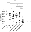
Treatment of MRSA ATCC 43300 K-wire-associated infection with different Sb-1 and daptomycin formulations in G. mellonella larvae. After infection with MRSA ATCC 43300 (105 CFUs) larvae were treated with (i) Sb-1 (107 PFUs) alone, (ii) daptomycin (4 mg/kg) alone and staggered administration of (iii) Sb-1 (107 PFUs)/PBS, (iv) PBS/daptomycin (4 mg/kg), and (v) Sb-1 (107 PFUs)/daptomycin (4 mg/kg). Statistical differences in bacterial count per K-wire obtained from different treatment groups were determined by the Kruskal–Wallis test using Dunn correction (* p ≤ 0.05; ** p ≤ 0.01; *** p ≤ 0.001; **** p < 0.0001). Data obtained from three independent experiments are reported.
Figure 5
Figure 5
Efficacy of Sb-1 in preventing MRSA ATCC 43300 K-wire colonization in a G. mellonella model of implant-associated infection. Sb-1 (107 PFUs) and vancomycin (10 mg/kg) were injected into larvae before the infection with MRSA ATCC 43300 (105 CFUs). The graphs show (a) CFU number of K-wires explanted 2 days post-infection from larvae pretreated with either Sb-1 or vancomycin; (b) CFU/mL number from hemolymph collected 2 days post-infection from larvae pretreated with either Sb-1 or vancomycin; (c) CFU number of K-wires explanted 5 days post-infection from larvae pretreated with either Sb-1 or vancomycin; (d) CFU number from hemolymph collected 5 days post-infection from larvae pretreated with either Sb-1 or vancomycin. Statistical differences in bacterial count per K-wire and bacterial count per mL of hemolymph obtained from different pre-treatment groups were determined by the Kruskal–Wallis test using Dunn correction (ns p > 0.05; ** p ≤ 0.01; *** p ≤ 0.001; **** p < 0.0001, ns = not significant). Data obtained from two independent experiments are reported.
Figure 6
Figure 6
Schematic representation of the experimental model for the establishment of implant-associated infection in G. mellonella larvae. (A) Evaluation of the optimum infectious dose of MRSA ATCC 43300 able to colonize K-wires implanted in larvae. (B) Sb-1 stability in hemolymph of G. mellonella larvae and kinetic of phage inactivation.
Figure 7
Figure 7
Schematic representation of the procedure used in the experimental model for the evaluation of the Sb-1 in vivo activity in treating (A) and preventing (B) implanted K-wire colonization by methicillin-resistant S. aureus ATCC 43300.

References

    1. Caldara M., Belgiovine C., Secchi E., Rusconi R. Environmental, Microbiological, and Immunological Features of Bacterial Biofilms Associated with Implanted Medical Devices. Clin. Microbiol. Rev. 2022;35:e0022120. doi: 10.1128/cmr.00221-20. - DOI - PMC - PubMed
    1. Arciola C.R., An Y.H., Campoccia D., Donati M.E., Montanaro L. Etiology of Implant Orthopedic Infections: A Survey on 1027 Clinical Isolates. Int. J. Artif. Organs. 2005;28:1091–1100. doi: 10.1177/039139880502801106. - DOI - PubMed
    1. Ricciardi B.F., Muthukrishnan G., Masters E., Ninomiya M., Lee C.C., Schwarz E.M. Staphylococcus Aureus Evasion of Host Immunity in the Setting of Prosthetic Joint Infection: Biofilm and Beyond. Curr. Rev. Musculoskelet. Med. 2018;11:389–400. doi: 10.1007/s12178-018-9501-4. - DOI - PMC - PubMed
    1. Lister J.L., Horswill A.R. Staphylococcus Aureus Biofilms: Recent Developments in Biofilm Dispersal. Front. Cell. Infect. Microbiol. 2014;4:178. doi: 10.3389/fcimb.2014.00178. - DOI - PMC - PubMed
    1. Osmon D.R., Berbari E.F., Berendt A.R., Lew D., Zimmerli W., Steckelberg J.M., Rao N., Hanssen A., Wilson W.R. Diagnosis and Management of Prosthetic Joint Infection: Clinical Practice Guidelines by the Infectious Diseases Society of Americaa. Clin. Infect. Dis. 2013;56:e1–e25. doi: 10.1093/cid/cis803. - DOI - PubMed

MeSH terms

LinkOut - more resources