

Mechanisms of Susceptibility and Resilience to PTSD: Role of Dopamine Metabolism and BDNF Expression in the Hippocampus
- PMID: 36498900
- PMCID: PMC9737079
- DOI: 10.3390/ijms232314575
Mechanisms of Susceptibility and Resilience to PTSD: Role of Dopamine Metabolism and BDNF Expression in the Hippocampus
Abstract
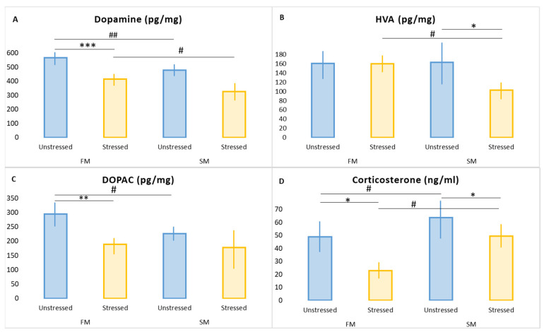
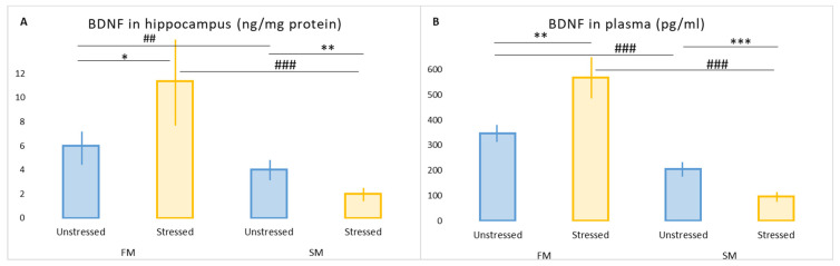
Susceptibility and resilience to post-traumatic stress disorder (PTSD) are recognized, but their mechanisms are not understood. Here, the hexobarbital sleep test (HST) was used to elucidate mechanisms of PTSD resilience or susceptibility. A HST was performed in rats 30 days prior to further experimentation. Based on the HST, the rats were divided into groups: (1) fast metabolizers (FM; sleep duration < 15 min); (2) slow metabolizers (SM; sleep duration ≥ 15 min). Then the SM and FM groups were subdivided into stressed (10 days predator scent, 15 days rest) and unstressed subgroups. Among stressed animals, only SMs developed experimental PTSD, and had higher plasma corticosterone (CORT) than stressed FMs. Thus, resilience or susceptibility to PTSD was consistent with changes in glucocorticoid metabolism. Stressed SMs had a pronounced decrease in hippocampal dopamine associated with increased expressions of catecholamine-O-methyl-transferase and DA transporter. In stressed SMs, a decrease in monoaminoxidase (MAO) A was associated with increased expressions of hippocampal MAO-A and MAO-B. BDNF gene expression was increased in stressed FMs and decreased in stressed SMs. These results demonstrate relationships between the microsomal oxidation phenotype, CORT concentration, and anxiety, and they help further the understanding of the role of the liver−brain axis during PTSD.
Keywords: BDNF; COMT; MAO; corticosterone; dopamine; hexobarbital sleep test; post-traumatic stress disorder.
Conflict of interest statement
The authors declare no conflict of interest. The funders had no role in the design of the study; in the collection, analyses, or interpretation of data; in the writing of the manuscript; or in the decision to publish the results.
Figures
Similar articles
-
Hexobarbital Sleep Test for Predicting the Susceptibility or Resistance to Experimental Posttraumatic Stress Disorder.Int J Mol Sci. 2020 Aug 17;21(16):5900. doi: 10.3390/ijms21165900. Int J Mol Sci. 2020. PMID: 32824478 Free PMC article.
-
High and low anxiety phenotypes in a rat model of complex post-traumatic stress disorder are associated with different alterations in regional brain monoamine neurotransmission.Psychoneuroendocrinology. 2020 Jul;117:104691. doi: 10.1016/j.psyneuen.2020.104691. Epub 2020 Apr 23. Psychoneuroendocrinology. 2020. PMID: 32361171
-
Individual susceptibility or resistance to posttraumatic stress disorder-like behaviours.Behav Brain Res. 2020 May 27;386:112591. doi: 10.1016/j.bbr.2020.112591. Epub 2020 Mar 16. Behav Brain Res. 2020. PMID: 32194190
-
Hypothalamic-pituitary-adrenal axis activity in post-traumatic stress disorder and cocaine use disorder.Stress. 2020 Nov;23(6):638-650. doi: 10.1080/10253890.2020.1803824. Epub 2020 Aug 24. Stress. 2020. PMID: 32835581 Review.
-
[Posttraumatic stress disorder (PTSD) as a consequence of the interaction between an individual genetic susceptibility, a traumatogenic event and a social context].Encephale. 2012 Oct;38(5):373-80. doi: 10.1016/j.encep.2011.12.003. Epub 2012 Jan 24. Encephale. 2012. PMID: 23062450 Review. French.
Cited by
-
Limited Cheese Intake Paradigm Replaces Patterns of Behavioral Disorders in Experimental PTSD: Focus on Resveratrol Supplementation.Int J Mol Sci. 2023 Sep 20;24(18):14343. doi: 10.3390/ijms241814343. Int J Mol Sci. 2023. PMID: 37762647 Free PMC article.
-
Resistance to Resveratrol Treatment in Experimental PTSD Is Associated with Abnormalities in Hepatic Metabolism of Glucocorticoids.Int J Mol Sci. 2023 May 26;24(11):9333. doi: 10.3390/ijms24119333. Int J Mol Sci. 2023. PMID: 37298287 Free PMC article.
-
Resveratrol: A Multifaceted Guardian against Anxiety and Stress Disorders-An Overview of Experimental Evidence.Nutrients. 2024 Aug 26;16(17):2856. doi: 10.3390/nu16172856. Nutrients. 2024. PMID: 39275174 Free PMC article. Review.
-
Theorizing that Psychedelic Assisted Therapy May Play a Role in the Treatment of Trauma-Induced Personality Disorders.J Addict Psychiatry. 2024;8(2):161-165. Epub 2024 Nov 15. J Addict Psychiatry. 2024. PMID: 39634920 Free PMC article.
References
-
- Tseilikman V., Komelkova M., Kondashevskaya M.V., Manukhina E., Downey H.F., Chereshnev V., Chereshneva M., Platkovskii P., Goryacheva A., Pashkov A., et al. A rat model of post-traumatic stress syndrome causes phenotype-associated morphological changes and hypofunction of the adrenal gland. Int. J. Mol. Sci. 2021;22:13235. doi: 10.3390/ijms222413235. - DOI - PMC - PubMed
-
- Manukhina E.B., Tseilikman V.E., Komelkova M.V., Lapshin M.S., Goryacheva A.V., Kondashevskaya M.V., Mkhitarov V.A., Lazuko S.S., Tseilikman O.B., Sarapultsev A.P., et al. Cardiac injury in rats with experimental posttraumatic stress disorder and mechanisms of its limitation in experimental posttraumatic stress disorder-resistant rats. J. Appl. Physiol. 2021;130:759–771. doi: 10.1152/japplphysiol.00694.2019. - DOI - PubMed
MeSH terms
Substances
Grants and funding
LinkOut - more resources
Full Text Sources
Medical
Miscellaneous
