Case Reports
. 2022 Nov 24;23(23):14643.
doi: 10.3390/ijms232314643.

Distant Recurrence of a Cerebral Cavernous Malformation in the Vicinity of a Developmental Venous Anomaly: Case Report of Local Oxy-Inflammatory Events

Affiliations
Case Reports

Distant Recurrence of a Cerebral Cavernous Malformation in the Vicinity of a Developmental Venous Anomaly: Case Report of Local Oxy-Inflammatory Events

Andrea Bianconi et al. Int J Mol Sci. .

Abstract

Background: Cerebral cavernous malformations (CCMs) are a major type of cerebrovascular lesions of proven genetic origin that occur in either sporadic (sCCM) or familial (fCCM) forms, the latter being inherited as an autosomal dominant condition linked to loss-of-function mutations in three known CCM genes. In contrast to fCCMs, sCCMs are rarely linked to mutations in CCM genes and are instead commonly and peculiarly associated with developmental venous anomalies (DVAs), suggesting distinct origins and common pathogenic mechanisms.

Case report: A hemorrhagic sCCM in the right frontal lobe of the brain was surgically excised from a symptomatic 3 year old patient, preserving intact and pervious the associated DVA. MRI follow-up examination performed periodically up to 15 years after neurosurgery intervention demonstrated complete removal of the CCM lesion and no residual or relapse signs. However, 18 years after surgery, the patient experienced acute episodes of paresthesia due to a distant recurrence of a new hemorrhagic CCM lesion located within the same area as the previous one. A new surgical intervention was, therefore, necessary, which was again limited to the CCM without affecting the pre-existing DVA. Subsequent follow-up examination by contrast-enhanced MRI evidenced a persistent pattern of signal-intensity abnormalities in the bed of the DVA, including hyperintense gliotic areas, suggesting chronic inflammatory conditions.

Conclusions: This case report highlights the possibility of long-term distant recurrence of hemorrhagic sCCMs associated with a DVA, suggesting that such recurrence is secondary to focal sterile inflammatory conditions generated by the DVA.

Keywords: cerebral cavernous malformation (CCM); cerebrovascular diseases; developmental venous anomaly (DVA); oxidative stress; sterile inflammation.


Conflict of interest statement

The authors declare no conflict of interest.

Figures

Figure 1
Figure 1
First preoperative T1 contrast-enhanced MRI showing a hemorrhagic cavernoma in the vicinity of a voluminous DVA, with its typical caput medusae sign, in the right frontal lobe: (A) axial view; (B) coronal view.
Figure 2
Figure 2
(A) Axial MRI view of the patient’s brain, showing the presence of a newly formed hemorrhagic CCM lesion close to the pre-existing DVA. (B) Postoperative axial MRI demonstrating complete removal of the newly formed CCM lesion.
Figure 3
Figure 3
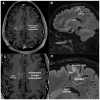
Contrast-enhanced MRI follow-up examination of the patient’s brain at 1 year after CCM surgery. (A,C) T1-weighted axial image. (B,D) T2-FLAIR (fluid-attenuated inversion recovery) sagittal image. Contrast-enhanced images were obtained after injection of gadolinium as nontoxic paramagnetic contrast enhancement agent. (C,D) Magnified images of panels (A,B), respectively. Notice the presence of signal-intensity abnormalities in the bed of the DVA, including point-like contrast enhancements and hyperintense gliotic areas.
Figure 4
Figure 4
Graphical representation of focal sterile oxy-inflammatory mechanisms potentially implicated in the formation and recurrence of a sporadic cerebral cavernous malformation (sCCM) in the drainage territory of a developmental venous anomaly (DVA). Sterile oxy-inflammatory and pro-angiogenic factors, including DAMPs, inflammatory cytokines, ROS, and VEGF, can be released by damaged neurovascular tissues and activated glial cells as a consequence of age-related increased abnormalities in the angioarchitectural and hemodynamic features of a DVA, as well as by other injuries that can contribute to focal gliosis in the adjacent brain parenchyma, such as radiation exposure, neurodegeneration, stroke, and hypoxia. In turn, such factors can induce aberrant oxy-inflammatory signaling pathways in local brain capillary endothelial cells through the activation of multivalent platforms of distinct but interrelated cell surface receptors and enzymes, including interleukin receptors (ILR), VEGF receptors (VEGFR), Toll-like receptors (TLR4), and NADPH oxidases (NOX), which eventually result in abnormal oxy-inflammatory and proangiogenic responses, thereby affecting the BBB stability and permeability, and enabling the focal onset of CCM lesions. In particular, these effects are mediated by multiple redox-dependent mechanisms, including NOX-mediated ROS production, upregulation of PI3K/Akt and MAPK signaling, modulation of major redox-sensitive transcription factors, such as FoxO1, NF-κB, c-Jun, and Nrf2, and mTOR activation-dependent downregulation of autophagy. Eventually, these multiple and interconnected mechanisms lead to abnormal adaptive responses that impair microvessel barrier function and increase their sensitivity to local oxy-inflammatory insults. Notably, these pathological effects can be further facilitated by gain-of-function mutations in genes involved in the PI3K/Akt and MAPK pathways, which have indeed been identified in surgical specimens of sCCM lesions (see Section 3 for further details). Abbreviations: AJ, adherens junction; BBB, blood–brain barrier; COX-2, cyclooxygenase-2; DAMPs, damage-associated molecular patterns; FoxO1, forkhead box O1; ILR, interleukin receptor; JNK, c-Jun N-terminal kinase; MAPK, mitogen-activated protein kinase; mTOR, mechanistic target of rapamycin; NF-κB, nuclear factor-κB; NOX, NADPH oxidases; Nrf2, nuclear factor erythroid 2-related factor 2; PI3K, phosphoinositide 3-kinase; ROS, reactive oxygen species; TJ, tight junctions; TLR4, Toll-like receptor 4; VEGF, vascular endothelial growth factor; VEGFR, VEGF receptor.

References

    1. Batra S., Lin D., Recinos P.F., Zhang J., Rigamonti D. Cavernous malformations: Natural history, diagnosis and treatment. Nat. Rev. Neurol. 2009;5:659–670. doi: 10.1038/nrneurol.2009.177. - DOI - PubMed
    1. Flemming K.D. Incidence, Prevalence, and Clinical Presentation of Cerebral Cavernous Malformations. Methods Mol. Biol. 2020;2152:27–33. doi: 10.1007/978-1-0716-0640-7_2. - DOI - PubMed
    1. Mabray M., Hart B. Clinical Imaging of Cerebral Cavernous Malformations: Computed Tomography and Magnetic Resonance Imaging. Methods Mol. Biol. 2020;2152:85–96. doi: 10.1007/978-1-0716-0640-7_7. - DOI - PubMed
    1. Ene C., Kaul A., Kim L. Natural history of cerebral cavernous malformations. Handb. Clin. Neurol. 2017;143:227–232. doi: 10.1016/B978-0-444-63640-9.00021-7. - DOI - PubMed
    1. Choquet H., Pawlikowska L., Lawton M.T., Kim H. Genetics of cerebral cavernous malformations: Current status and future prospects. J. Neurosurg. Sci. 2015;59:211–220. - PMC - PubMed

Publication types

MeSH terms

LinkOut - more resources