. 2022 Nov 24;23(23):14657.
doi: 10.3390/ijms232314657.

Immunoproteasome Inhibition Ameliorates Aged Dystrophic Mouse Muscle Environment

Affiliations

Immunoproteasome Inhibition Ameliorates Aged Dystrophic Mouse Muscle Environment

Luana Tripodi et al. Int J Mol Sci. .

Abstract

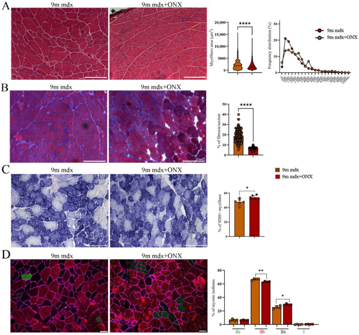
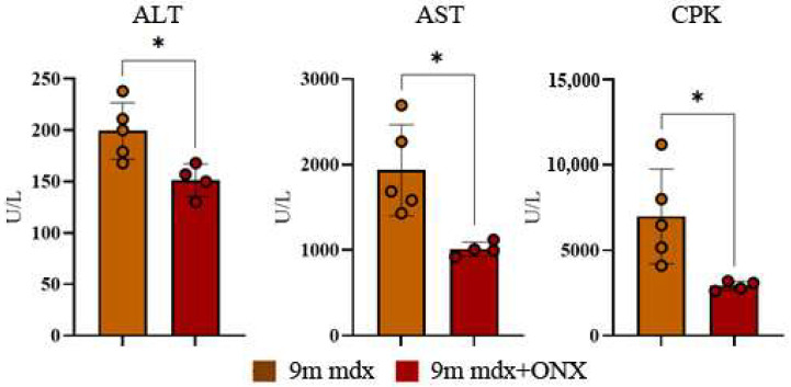
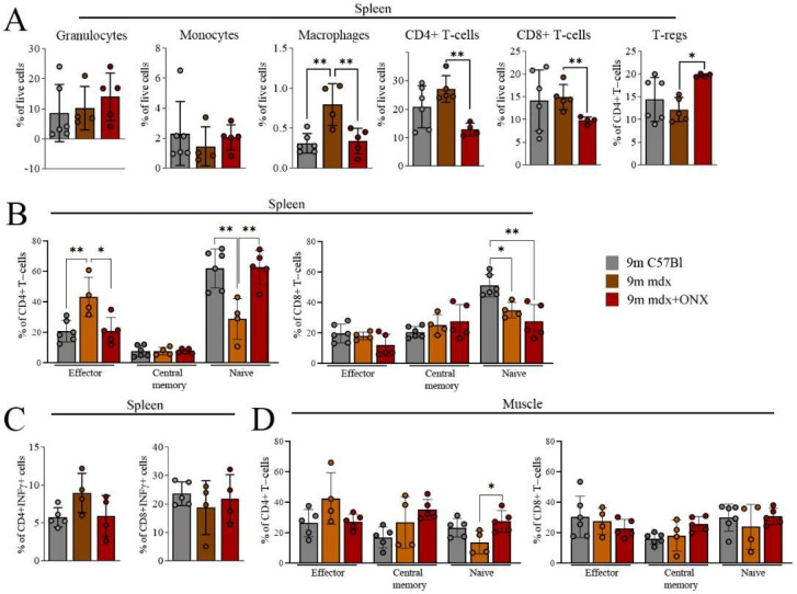
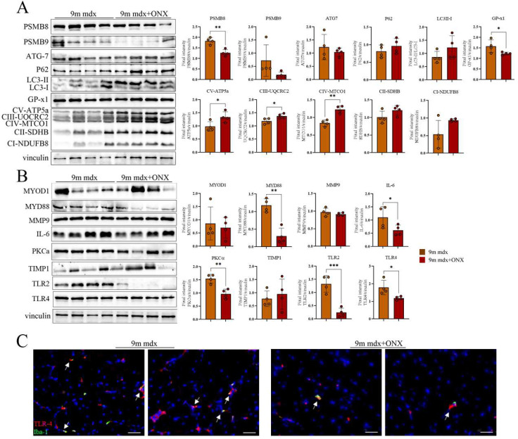
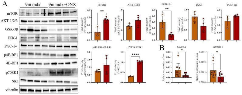
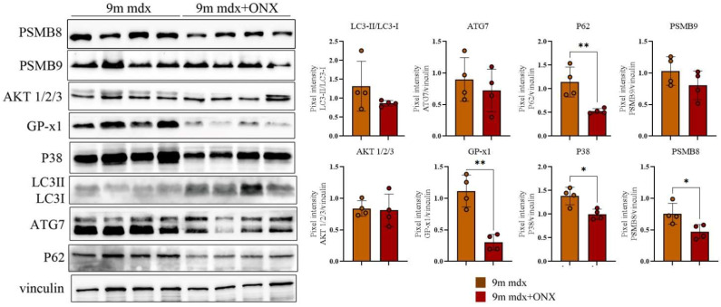
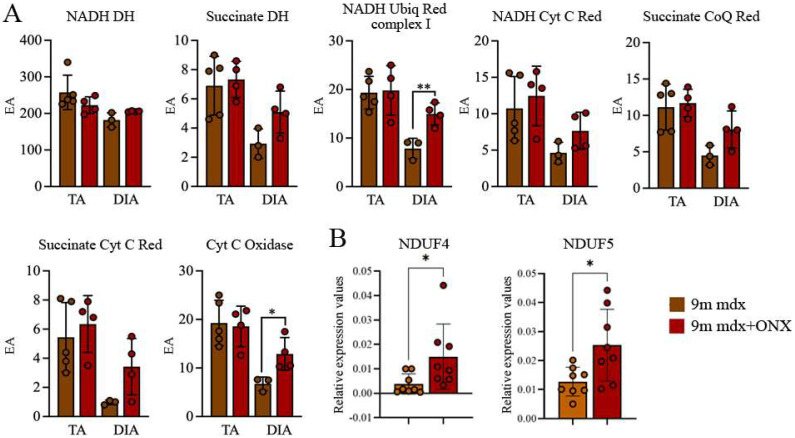
Muscle wasting is a major pathological feature observed in Duchenne muscular dystrophy (DMD) and is the result of the concerted effects of inflammation, oxidative stress and cell senescence. The inducible form of proteasome, or immunoproteasome (IP), is involved in all the above mentioned processes, regulating antigen presentation, cytokine production and immune cell response. IP inhibition has been previously shown to dampen the altered molecular, histological and functional features of 3-month-old mdx mice, the animal model for DMD. In this study, we described the role of ONX-0914, a selective inhibitor of the PSMB8 subunit of immunoproteasome, in ameliorating the pathological traits that could promote muscle wasting progression in older, 9-month-old mdx mice. ONX-0914 reduces the number of macrophages and effector memory T cells in muscle and spleen, while increasing the number of regulatory T cells. It modulates inflammatory markers both in skeletal and cardiac muscle, possibly counteracting heart remodeling and hypertrophy. Moreover, it buffers oxidative stress by improving mitochondrial efficiency. These changes ultimately lead to a marked decrease of fibrosis and, potentially, to more controlled myofiber degeneration/regeneration cycles. Therefore, ONX-0914 is a promising molecule that may slow down muscle mass loss, with relatively low side effects, in dystrophic patients with moderate to advanced disease.

Keywords: aging; immunoproteasome; inflammation; muscle mass; sarcopenia.


Conflict of interest statement

The authors declare no conflict of interest.

Figures

Figure 1
Figure 1
Histological and immunofluorescence analysis of tibialis anterior section of 9m mdx and 9m mdx+ONX mice. (A) Representative H&E staining and quantification of myofiber area and relative frequency of myofiber cross-sectional areas (CSA) expressed as frequency distribution of TA muscles of 9m mdx and 9m mdx+ONX mice (n = 10 images for animal group). Scale bar: 200 μm. (B) Quantification of fibrotic area in TA muscles of 9m mdx and 9m mdx+ONX mice (n = 10 images for animal group). (C) Representative SDH staining and quantification of percentage of SDH+ myofibers in TA of 9m mdx and 9m mdx+ONX mice (n = 3 for all groups). Scale bar: 200 μm. (D) Representative images of immunofluorescent staining showing distribution of myosin heavy chain (MyHC) isoforms (Type I, Type IIa, Type IIx and Type IIb) (n = 8 images were analyzed for each mouse). Graph portrays the percentage of myofibers expressing different MyHC isoforms. Scale bar: 50 μm. For morphometric analysis, images were quantified with Image J software. (* p < 0.05, ** p <0.01, **** p < 0.0001, with unpaired t-test).
Figure 2
Figure 2
Serum ALT, AST and CPK levels in 9m mdx and 9m mdx+ONX mice. ALT, AST and CPK were measured in blood serum drawn from 9m mdx (n = 5) and 9m mdx+ONX mice (n = 4). Data are expressed as means ± SD of n = 4–5 9m mdx and 9m mdx+ONX mice (* p < 0.05, with unpaired t-test).
Figure 3
Figure 3
FACS analysis of immune cell population from spleen and skeletal muscles of 9m C57Bl, 9m mdx and 9m mdx+ONX mice. FACS analysis was performed on spleen and muscle homogenates from C57Bl, 9m mdx and 9m mdx+ONX mice. Panels for spleen show (A) percentages of granulocytes (Ly6g+), monocytes (Ly6c+), macrophages (F4/80+CD11b+), CD4+ and CD8+ T cells, and Tregs (FOXP3+ on CD4+ T-cell gate); (B) frequency of effector (CD62L−CD44+), central memory (CD62L+CD44+) and naïve (CD62L+CD44−) T cells within the CD4+ or CD8+ populations; (C) frequency of CD4+ and CD8+ IFNγ-producing cells. (D) Panel for muscle displays frequency of effector (CD62L−CD44+), central memory (CD62L+CD44+) and naïve (CD62L+CD44) T-cells within the CD4+ or CD8+ populations. Data are expressed as means ± SD of n = 2 independent experiments with n = 6 C57Bl mice and n = 4–5 9m mdx and 9m mdx+ONX mice (* p < 0.05, ** p < 0.01, with ordinary one-way ANOVA, Tukey’s multiple comparison test).
Figure 4
Figure 4
Anti-inflammatory and fibrotic effects of ONX-0194 on skeletal muscle of 9m mdx mice. Cropped images of representative WB showing the expression of proteins involved in (A) immunoproteasome, autophagy, oxidative phosphorylation, and (B) fibrosis and inflammation in skeletal muscles from 9m mdx mice treated with ONX-0914 versus untreated mice. Densitometric analyses of protein expression is shown as a ratio on vinculin. (C) Fluorescent staining of TLR-4 (in red) and Iba-1 (in green) while nuclei are in DAPI (blu) (scale bar: 50 μm) in TA of 9m mdx and 9m mdx+ONX. Macrophages expressing TLR-4 are evidenced by white arrows. Data are expressed as means ± SD of n = 3 independent experiments with n = 4 mice for animal groups (* p < 0.05, ** p < 0.01, *** p < 0.001, with unpaired t-test).
Figure 5
Figure 5
Evaluation of muscle wasting in 9m mdx and 9m mdx+ONX mice. Cropped images of representative WB showing (A) the expression of proteins involved muscle wasting as GSK-3β, IKK-i and PGC-1α; mTOR/AKT signaling and its most-common downstream targets 4E-BP1 and SK in skeletal muscles from 9m mdx mice treated with ONX-0914 versus untreated mice. Densitometric analyses of protein expression is shown as ratio on vinculin or ratio phosphorylated/total isoforms. (B) RT-qPCR analysis of MuRF-1 and atrogin genes TA of 9m mdx and 9m mdx+ONX. Data are expressed as means ± SD of n = 3 independent experiments with n = 4 mice for animal groups (* p < 0.05, ** p < 0.01, **** p < 0.0001, with unpaired t-test).
Figure 6
Figure 6
Proteomic analysis of whole cardiac muscle lysates from 9m mdx and 9m mdx+ONX mice. Cropped images of representative WB showing the expression of proteins involved in autophagy, fibrosis and inflammation in cardiac tissues from 9m mdx mice treated with ONX-0914 versus untreated mice. Densitometric analyses of protein expression is shown as ratio on vinculin. Data are expressed as means ± SD of n = 3 independent experiments with n = 4 mice for animal groups (* p < 0.05, ** p < 0.01, with unpaired t-test).
Figure 7
Figure 7
Mitochondrial respiratory chain enzymatic activity in TA and DIA of 9m mdx and 9m mdx+ONX mice. (A) Effects of ONX-0914 treatment on the activity of muscle (TA and DIA) respiratory chain enzymes in 9m mdx and 9m mdx+ONX mice (EA: enzymatic activity; NADH DH: NADH dehydrogenase; Succinate DH: succinate dehydrogenase; NADH Ubiq Red complex 1: NADH ubiquinone reductase complex 1; NADH Cyt C Red: NADH cytochrome C reductase; Succinate CoQ Red: succinate CoQ reductase; Succinate Cyt C Red: succinate cytochrome C reductase; Cyt C oxidase: cytochrome C oxidase). (B) RT-qPCR analysis of NDUF4 and NDUF5 in DIA of 9m mdx and 9m mdx+ONX mice. Data are expressed as means ± SD of n = 2 independent experiments with n = 4–5 9m mdx and 9m mdx+ONX mice (* p < 0.05, ** p < 0.01, with unpaired t-test). ONX-0914 upregulates NADH ubiquinone 1 reductase and of cytochrome oxidase.

References

    1. Villalta S.A., Rosenberg A.S., Bluestone J.A. The immune system in Duchenne muscular dystrophy: Friend or foe. Rare Dis. 2015;3:e1010966. doi: 10.1080/21675511.2015.1010966. - DOI - PMC - PubMed
    1. Merlini L., Vagheggini A., Cocchi D. Sarcopenia and sarcopenic obesity in patients with muscular dystrophy. Front. Aging Neurosci. 2014;6:274. doi: 10.3389/fnagi.2014.00274. - DOI - PMC - PubMed
    1. Ubaida-Mohien C., Lyashkov A., Gonzalez-Freire M., Tharakan R., Shardell M., Moaddel R., Semba R.D., Chia C.W., Gorospe M., Sen R., et al. Discovery proteomics in aging human skeletal muscle finds change in spliceosome, immunity, proteostasis and mitochondria. eLife. 2019;8:e49874. doi: 10.7554/eLife.49874. - DOI - PMC - PubMed
    1. Sugihara H., Teramoto N., Nakamura K., Shiga T., Shirakawa T., Matsuo M., Ogasawara M., Nishino I., Matsuwaki T., Nishihara M., et al. Cellular senescence-mediated exacerbation of Duchenne muscular dystrophy. Sci. Rep. 2020;10:16385. doi: 10.1038/s41598-020-73315-6. - DOI - PMC - PubMed
    1. Brehm M.A., Kempen J.C., van der Kooi A.J., de Groot I.J., van den Bergen J.C., Verschuuren J.J., Niks E.H., Harlaar J. Age-related longitudinal changes in metabolic energy expenditure during walking in boys with Duchenne muscular dystrophy. PLoS ONE. 2014;9:e115200. doi: 10.1371/journal.pone.0115200. - DOI - PMC - PubMed