. 2022 Nov 24;23(23):14681.
doi: 10.3390/ijms232314681.

Optimal Intravenous Administration Procedure for Efficient Delivery of Canine Adipose-Derived Mesenchymal Stem Cells

Affiliations

Optimal Intravenous Administration Procedure for Efficient Delivery of Canine Adipose-Derived Mesenchymal Stem Cells

Yuyo Yasumura et al. Int J Mol Sci. .

Abstract

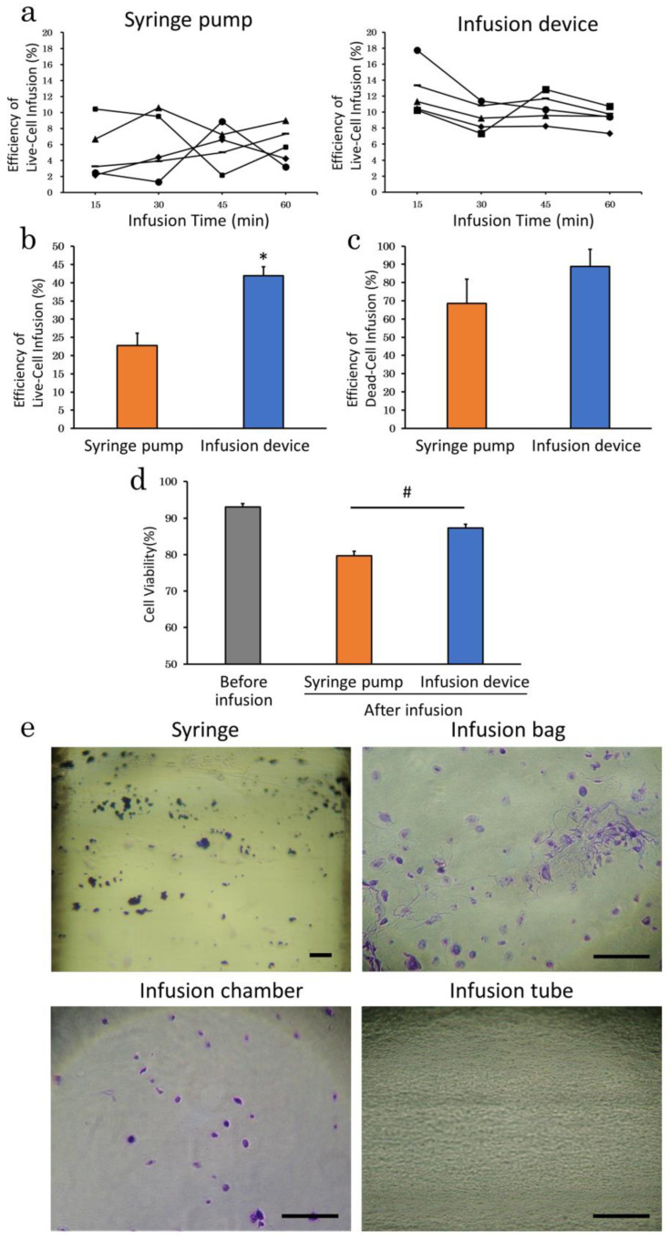
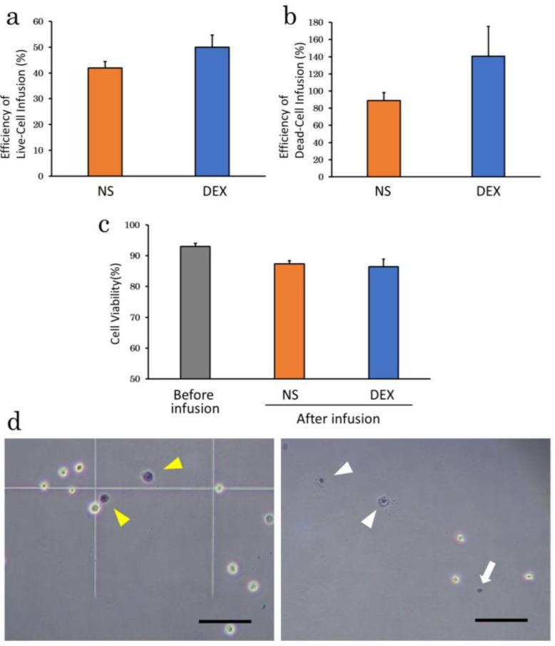
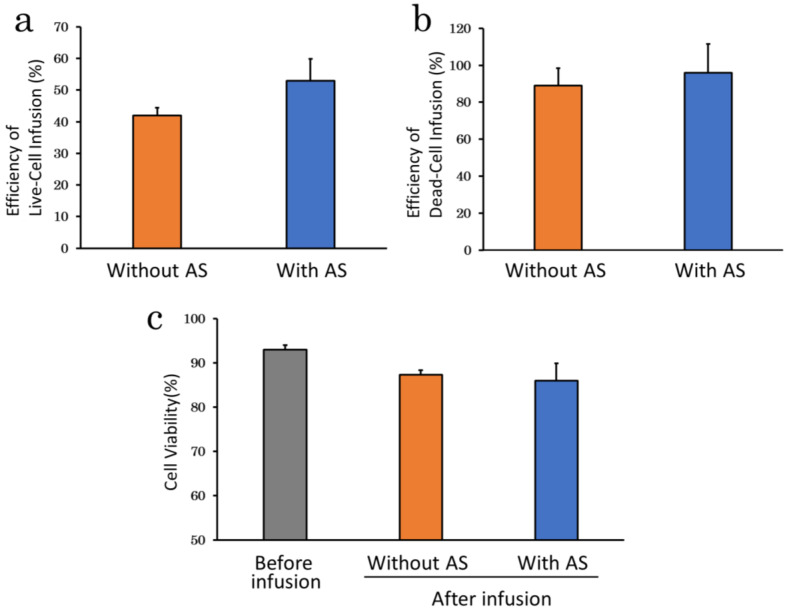
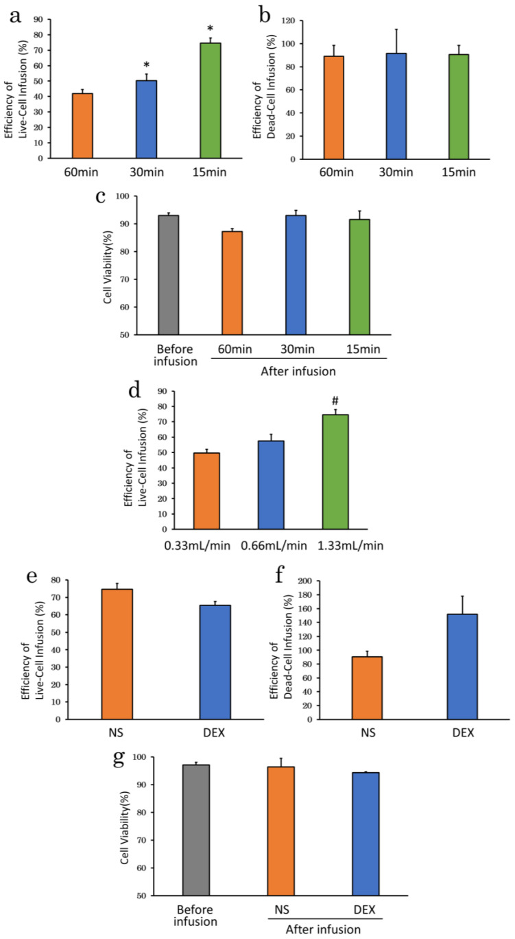
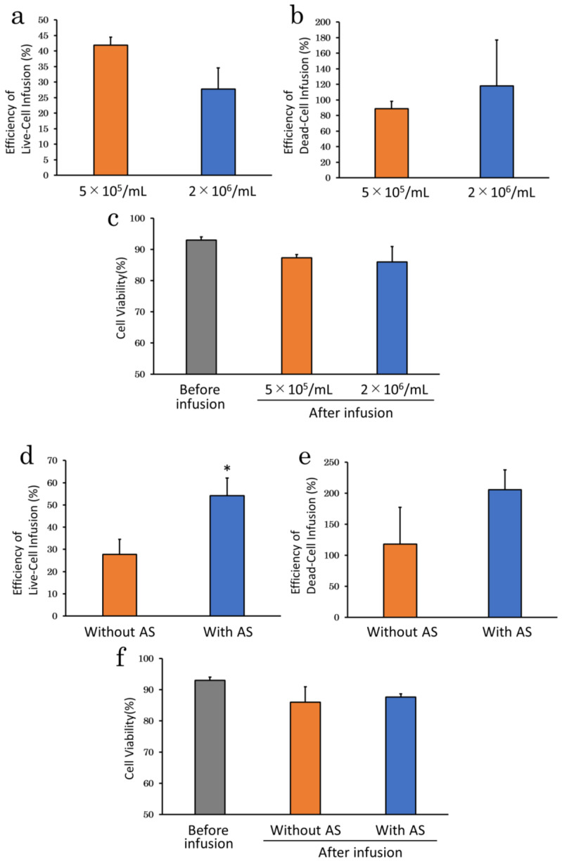
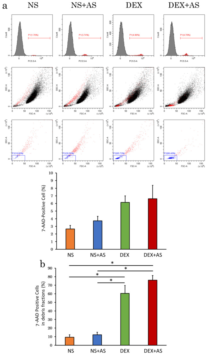
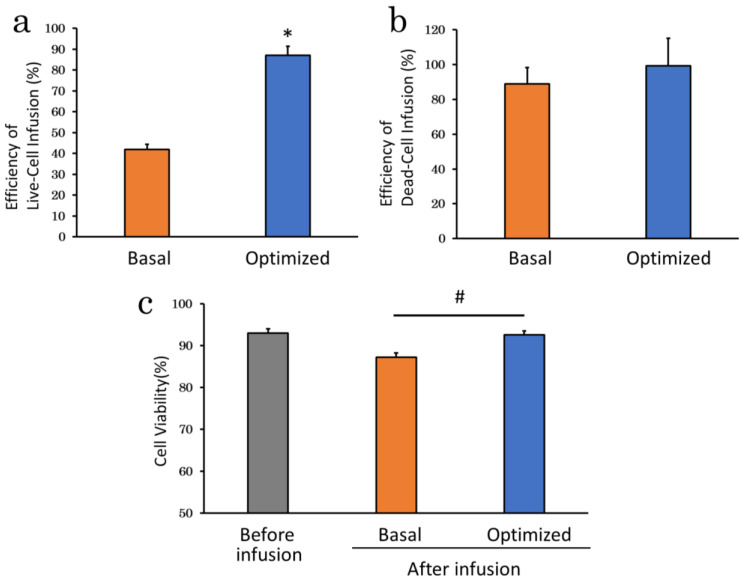
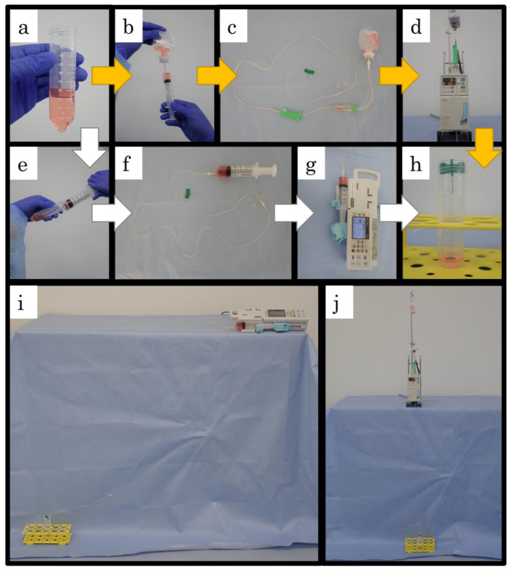
Mesenchymal stem cells (MSC) are currently being investigated for their therapeutic applications in a wide range of diseases. Although many studies examined peripheral venous administration of MSC, few have investigated the detailed intravenous administration procedures of MSC from their preparation until they enter the body. The current study therefore aimed to explore the most efficient infusion procedure for MSC delivery by preparing and infusing them under various conditions. Canine adipose-derived mesenchymal stem cells (cADSC) were infused using different infusion apparatuses, suspension solutions, allogenic serum supplementation, infusion time and rates, and cell densities, respectively. Live and dead cell counts were then assessed by manual measurements and flow cytometry. Efficiency of live- and dead-cell infusion and cell viability were calculated from the measured cell counts and compared under each condition. Efficiency of live-cell infusion differed significantly according to the infusion apparatus, infusion rate, and combination of cell density and serum supplementation. Cell viability after infusion differed significantly between the infusion apparatuses. The optimal infusion procedure resulting in the highest cell delivery and viability involved suspending cADSC in normal saline supplemented with 5% allogenic serum at a density of 5 × 10⁵ cells/mL, and infusing them using an automatic infusion device for 15 min. This procedure is therefore recommended as the standard procedure for the intravenous administration of ADSC in terms of cell-delivery efficiency.

Keywords: adipose-derived mesenchymal stem cells; canine; cell delivery; cell viability; intravenous administration.


Conflict of interest statement

The authors declare that there is no conflict of interest regarding the publication of this paper.

Figures

Figure 1
Figure 1
Differentiation of cADSC into three lineages. (a), Adipogenic differentiation identified by Oil Red O staining; (b), Osteogenic differentiation identified by Alizarin Red staining; (c), Chondrogenic differentiation identified by Alcian Blue. Bar = 100 μm.
Figure 2
Figure 2
Infusion apparatus experiments. (a), Efficiency of live-cell infusion every 15 min using syringe pump (left panel) and infusion device (right panel). Each broken line represents the results of five independent experiments (n = 5); (b), Efficiency of live-cell infusion over 60 min. The infusion device increased efficiency of live-cell infusion approximately two-fold compared with the syringe pump; (c), Efficiency of dead-cell infusion over 60 min; (d), Cell viability after 60 min infusion. The cell viability using an infusion device was significantly higher than using a syringe pump; (e), Adherent cells in the apparatuses stained by Giemsa staining. cADSC adhered to various parts of the infusion apparatuses. No cell adhesion was observed in the tubes. * p < 0.01 vs. syringe pump. # p < 0.01, between groups. Bar = 200 μm.
Figure 3
Figure 3
Efficiency of cell infusion and cell viability in different suspension solutions. (a), Efficiency of live-cell infusion for NS and DEX. There were no significant differences, but efficiency of live-cell infusion tended to be higher in DEX; (b), Efficiency of dead-cell infusion for NS and DEX. Efficiency of dead-cell infusion also tended to be higher in DEX; (c), Cell viability after infusion for NS and DEX showed a slightly lower in DEX; (d), Microscope images of dead cADSC suspended in NS (left panel) or DEX (right panel). Trypan blue-stained dead cells suspended in NS were comparable in size and morphology to live cells (left panel; yellow arrowheads). Small concentrated Trypan Blue-stained dead cells (right panel; white arrows) and cells that appeared to be bursting (right panel; white arrowheads) were observed in DEX. NS: normal saline; DEX: 5% dextrose. Bar = 100 μm.
Figure 4
Figure 4
Differences in efficiency of cell infusion and cell viability with and without AS. (a), Efficiency of live-cell infusion with and without AS. There were no significant differences, but efficiency of live-cell infusion tended to be higher with AS; (b), Efficiency of dead-cell infusion with and without AS. Efficiency of dead-cell infusion also tended to be higher with AS; (c), Cell viability with and without AS. Supplementation of AS slightly decreased in viability. AS: allogenic serum.
Figure 5
Figure 5
Infusion time experiments. (a), Efficiency of live-cell infusion for 15, 30, and 60 min infusions. Efficiency of live-cell infusion was significantly higher for 15 and 30 min than for 60 min; (b), Efficiency of dead-cell infusion did not differ among the three time groups; (c), Cell viability after infusion tended to be higher at 15 and 30 min; (d), Differences in efficiency of live-cell infusion by infusion rate. Faster infusion rates tended to result in higher efficiency of live-cell infusion; (e), Efficiency of live-cell infusion after 15 min infusion with different suspension solutions. In contrast to infusion over 60 min, efficiency of live-cell infusion tended to be better in NS; (f), Efficiency of dead-cell infusion after 15 min infusion with different suspension solutions. Efficiency of dead-cell infusion was high at DEX, exceeding 100%; (g), Cell viability after 15 min infusion with different suspension solutions. * p < 0.01 vs. 60 min. # p < 0.05 vs. 0.33 mL/min. NS: normal saline; DEX: 5% dextrose.
Figure 6
Figure 6
Cell density experiments. (a), Efficiency of live-cell infusion at different cell densities. Higher cell density tended to result in lower efficiency of live-cell infusion; (b), Efficiency of dead-cell infusion at different cell densities. Higher cell density tended to result in higher efficiency of dead-cell infusion; (c), Cell viability at different cell densities. Higher cell density resulted in lower cell viability after infusion; (d), Effect of serum supplementation at high cell density. AS supplementation significantly improved efficiency of live-cell infusion at high cell density; (e), Effect of serum supplementation on efficiency of live-cell infusion at high cell densities; (f), Differences in cell viability after infusion with and without serum supplementation at high cell densities. * p < 0.05 vs. without AS. AS: allogenic serum.
Figure 7
Figure 7
Flow cytometry analysis of dead cells stained with 7-AAD. (a), Dot plots and histograms of all analyzed cells (top and middle rows), and dot plots of 7-AAD-positive cells gated by debris fraction (lower row). Although there was no significant difference, percentage of 7-AAD-positive cells were lower with NS than with DEX, and without than with AS (p = 0.05); (b), Suspension in DEX significantly increased 7-AAD-positive cells in the debris fraction. * p < 0.01, between groups. NS: normal saline; AS: allogenic serum; DEX: 5% dextrose.
Figure 8
Figure 8
Optimized infusion procedure. (a), Efficiency of live-cell infusion under optimized conditions. The optimized infusion procedure significantly improved efficiency of live-cell infusion compared with the basal condition, which were the best results in this study; (b), Efficiency of dead-cell infusion of infusion under optimized conditions. Efficiency of dead-cell infusion was slightly higher than the basal condition; (c), Cell viability of infusion under optimized conditions. The optimized infusion procedure significantly improved cell viability after infusion compared with the basal condition. * p < 0.01 vs. control. # p < 0.01, between groups.
Figure 9
Figure 9
Outline the infusion procedures. (a), Suspend 1 × 10⁷ cADSC in 20 mL NS to prepare a cell suspension with a density of 5 × 10⁵ cells/mL; (b), The prepared solution is collected in a 25 mL syringe with an 18G needle and transferred to an empty 50 mL infusion bag; (c), The cell-containing bag is connected to the infusion tube with a 21G winged needle; (d), The bag is placed in a drop-controlled automatic infusion device; (e), The prepared solution is collected directly in a 50 mL syringe with an 18G needle; (f), the cell-filled syringe is connected to an extension tube with a 21G winged needle; (g), the syringe was placed in a syringe pump; (h), The winged needle tip is placed in a conical tube and the infused cells were collected. The conical tubes are replaced every 15 min, mixed immediately by gentle inversion; (i), Overall view of an infusion using a syringe pump. The conical tube for collecting the flowing cell suspension is placed lower than the syringe pump and the extension tube is not deflected; (j), Overall view of an infusion using an infusion device. The cell suspensions in this series of photographs are stained red for photography to visualize suspension transfer. Yellow arrow: infusion procedure using an infusion device; White arrow: infusion procedure using a syringe pump.

Similar articles

Cited by

References

    1. Andrzejewska A., Dabrowska S., Lukomska B., Janowski M. Mesenchymal stem cells for neurological disorders. Adv. Sci. 2021;8:2002944. doi: 10.1002/advs.202002944. - DOI - PMC - PubMed
    1. Jorgensen C., Noël D. Mesenchymal stem cells in osteoarticular diseases. Regen. Med. 2011;6:44–51. doi: 10.2217/rme.11.80. - DOI - PubMed
    1. Bagno L., Hatzistergos K.E., Balkan W., Hare J.M. Mesenchymal stem cell-based therapy for cardiovascular disease: Progress and challenges. Mol. Ther. 2018;26:1610–1623. doi: 10.1016/j.ymthe.2018.05.009. - DOI - PMC - PubMed
    1. Markov A., Thangavelu L., Aravindhan S., Zekiy A.O., Jarahian M., Chartrand M.S., Pathak Y., Marofi F., Shamlou S., Hassanzadeh A. Mesenchymal stem/stromal cells as a valuable source for the treatment of immune-mediated disorders. Stem. Cell Res. Ther. 2021;12:192. doi: 10.1186/s13287-021-02265-1. - DOI - PMC - PubMed
    1. Dominici M., Blanc K.L., Mueller I., Slaper-Cortenbach I., Marini F., Krause D., Deans R., Keating A., Prockop D., Horwitz E. Minimal criteria for defining multipotent mesenchymal stromal cells. The international society for cellular therapy position statement. Cytotherapy. 2006;8:315–317. doi: 10.1080/14653240600855905. - DOI - PubMed

LinkOut - more resources