. 2022 Nov 24;23(23):14687.
doi: 10.3390/ijms232314687.

Fusion Cell Markers in Circulating Tumor Cells from Patients with High-Grade Ovarian Serous Carcinoma

Affiliations

Fusion Cell Markers in Circulating Tumor Cells from Patients with High-Grade Ovarian Serous Carcinoma

Anna Paula Carreta Ruano et al. Int J Mol Sci. .

Abstract

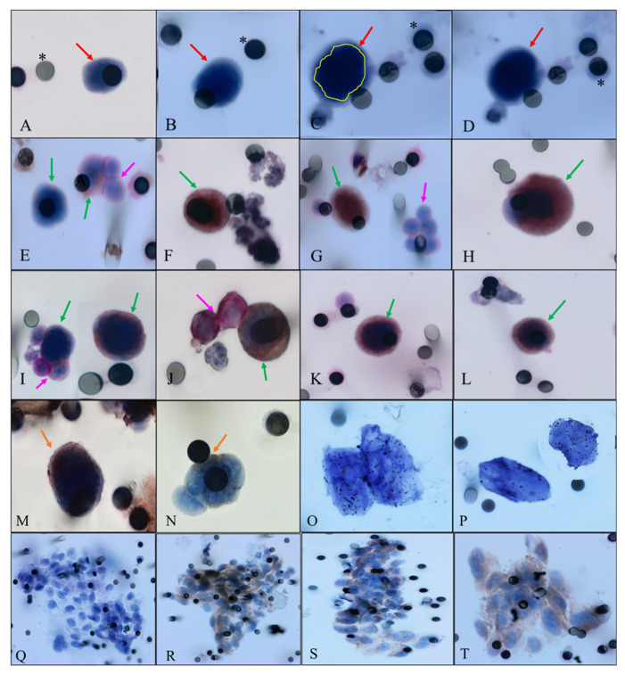
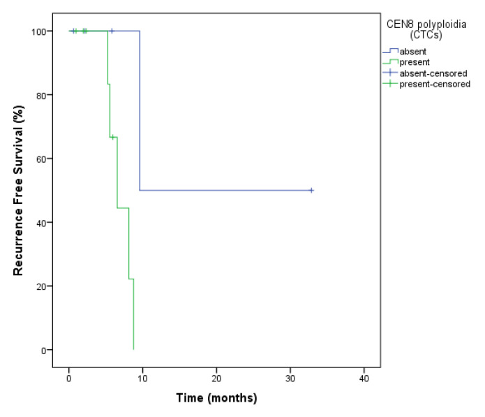
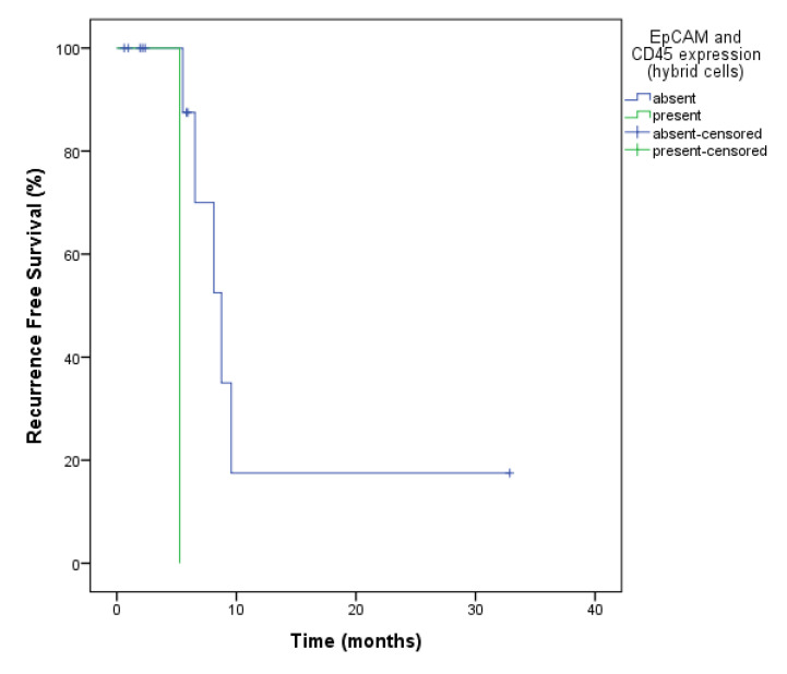
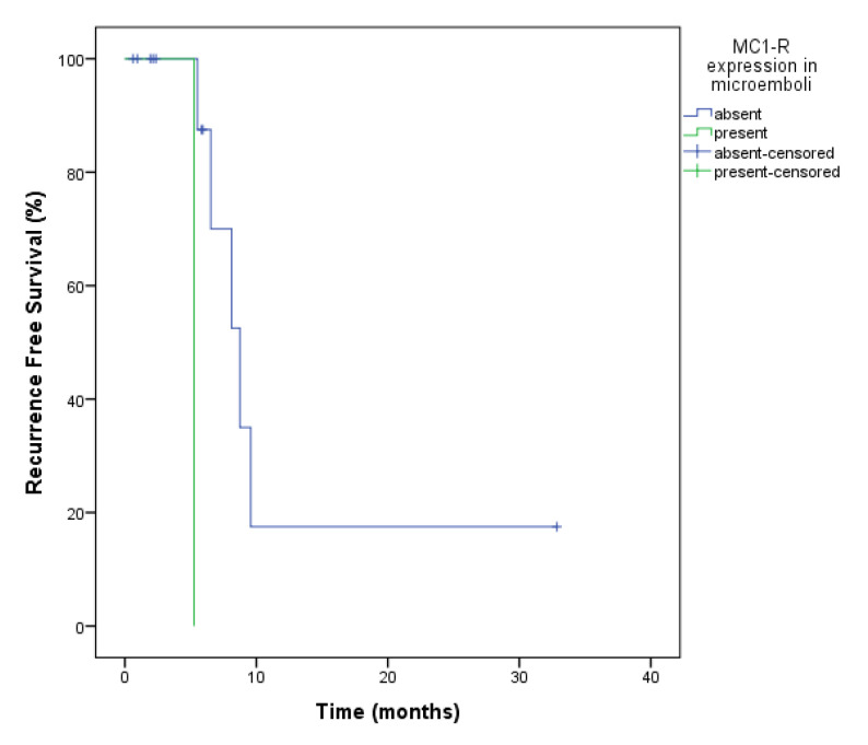
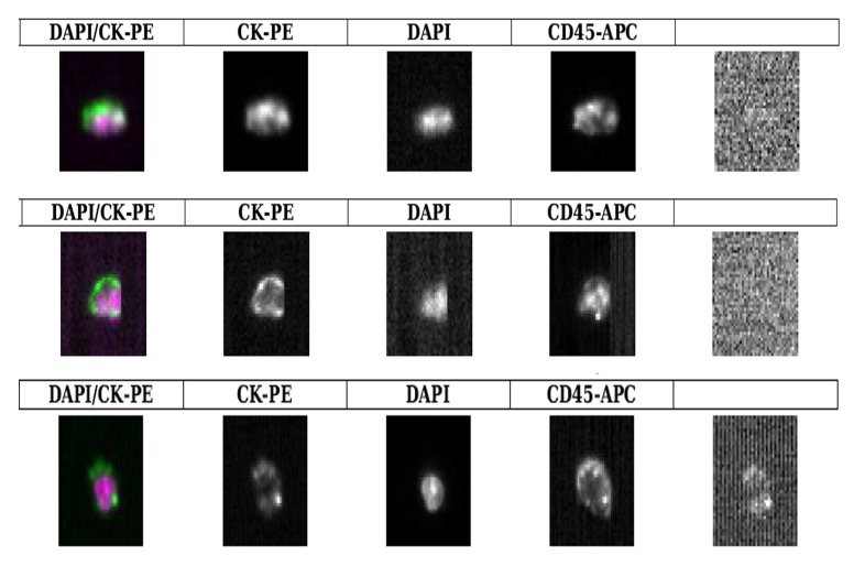
Cancer is primarily a disease in which late diagnosis is linked to poor prognosis, and unfortunately, detection and management are still challenging. Circulating tumor cells (CTCs) are a potential resource to address this disease. Cell fusion, an event discovered recently in CTCs expressing carcinoma and leukocyte markers, occurs when ≥2 cells become a single entity (hybrid cell) after the merging of their plasma membranes. Cell fusion is still poorly understood despite continuous evaluations in in vitro/in vivo studies. Blood samples from 14 patients with high-grade serous ovarian cancer (A.C. Camargo Cancer Center, São Paulo, Brazil) were collected with the aim to analyze the CTCs/hybrid cells and their correlation to clinical outcome. The EDTA collected blood (6 mL) from patients was used to isolate/identify CTCs/hybrid cells by ISET. We used markers with possible correlation with the phenomenon of cell fusion, such as MC1-R, EpCAM and CD45, as well as CEN8 expression by CISH analysis. Samples were collected at three timepoints: baseline, after one month (first follow-up) and after three months (second follow-up) of treatment with olaparib (total sample = 38). Fourteen patients were included and in baseline and first follow-up all patients showed at least one CTC. We found expression of MC1-R, EpCAM and CD45 in cells (hybrid) in at least one of the collection moments. Membrane staining with CD45 was found in CTCs from the other cohort, from the other center, evaluated by the CellSearch® system. The presence of circulating tumor microemboli (CTM) in the first follow-up was associated with a poor recurrence-free survival (RFS) (5.2 vs. 12.2 months; p = 0.005). The MC1-R expression in CTM in the first and second follow-ups was associated with a shorter RFS (p = 0.005). CEN8 expression in CTCs was also related to shorter RFS (p = 0.035). Our study identified a high prevalence of CTCs in ovarian cancer patients, as well as hybrid cells. Both cell subtypes demonstrate utility in prognosis and in the assessment of response to treatment. In addition, the expression of MC1-R and EpCAM in hybrid cells brings new perspectives as a possible marker for this phenomenon in ovarian cancer.

Keywords: CD45; cell fusion; circulating tumor cells; hybrid cells; in situ hybridization; ovarian cancer.


Conflict of interest statement

C.A.-P. received honoraria from Menarini. The remaining authors declare no conflict of interest relevant to the manuscript.

Figures

Figure 1
Figure 1
Representative images. Green arrows indicate hybrid cells, pink arrows represent leukocytes, red arrows represent CTCs, orange arrows indicate hybrids cells stained with CD45, yellow circle indicate cell nucleus and asterisks represent membrane pore. (A,B) CTCs without any staining, visualized with hematoxilin. (C,D)The same CTC without any staining, in (C), nuclei defined by yellow line. (E) Hybrid cells. One positive for MC1-R (brown membrane) and one in a microemboli, stained with CD45 with two leukocytes (stained with CD45, visualized by permanent red). (F) Hybrid cell positive for MC1-R (brown membrane). (G) Hybrid cell positive for EpCAM (brown membrane) and a cluster of leukocytes (stained with CD45, visualized by permanent red). (H) Hybrid cell double positive for EpCAM (brown membrane) and MC1-R (visualized by permanent red). (I) Two hybrid cells. One visualized alone double stained with EpCAM and CD45 (brown and red) and the other surrounded by three leukocytes stained with CD45 (visualized by permanent red). (J) Hybrid cell stained with MC1-R, visualized by DAB and two leukocytes (stained with CD45, visualized by permanent red). (K,L) Hybrid cells positive for MC1-R/CD45 (brown and red membrane). (M,N) Hybrids cells stained with CD45 in just one side of the cytoplasmatic membrane, visualized by DAB. (O,P) CTCs positive for CEN8, indicating the presence of polyploidy. (Q) Microemboli visualized with hematoxylin. (RT) Microemboli from patient positive for MC1-R (brown membrane). Images were taken at ×400 and ×600 magnification using a light microscope (Research System Microscope BX61—Olympus, Tokyo, Ja-pan) coupled to a digital camera (SC100—Olympus, Tokyo, Japan).
Figure 2
Figure 2
Recurrence-free survival analysis of ovarian cancer patients for CEN8 expression in CTCs (hybrid cells) at the baseline. The presence of polyploidy in the CEN8 showed a worse RFS (7.0 months vs. 21.21 months, p = 0.035) in relation to those who did not.
Figure 3
Figure 3
Recurrence-free survival analysis of ovarian cancer patients for EpCAM and CD45 expression in hybrid cells at the baseline and at second follow-up. The expression of this markers showed a worse RFS of 5.26 months vs. 12.2 (p = 0.005) in relation to those who did not.
Figure 4
Figure 4
Recurrence-Free Survival analysis of ovarian cancer patients for MC1-R expression in CTM. The presence of a microemboli with MC1-R expression at the second follow-up was also determinant for worse RFS (5.26 vs. 13.1 months; p = 0.008).
Figure 5
Figure 5
CTCs isolated from 3 different patients with locally advanced ovarian cancer with peritoneal carcinomatosis but no distant metastases (M0). These photos are representative of dual stained CTCs detected using the CellSearch® system: dual CK(+) CD45(+) nucleated cells after EpCAM-based enrichment step.

References

    1. Bray F., Msc M.L., Weiderpass E., Soerjomataram I. The ever-increasing importance of cancer as a leading cause of premature death worldwide. Cancer. 2021;127:3029–3030. doi: 10.1002/cncr.33587. - DOI - PubMed
    1. Sung H., Ferlay J., Siegel R.L., Laversanne M., Soerjomataram I., Jemal A., Bray F. Global Cancer Statistics 2020: GLOBOCAN Estimates of Incidence and Mortality Worldwide for 36 Cancers in 185 Countries. CA Cancer J. Clin. 2021;71:209–249. doi: 10.3322/caac.21660. - DOI - PubMed
    1. Instituto Nacional de Câncer-INCA INCA. Published 2022. [(accessed on 20 September 2022)]; Available online: https://www.inca.gov.br/tipos-de-cancer/cancer-de-ovario.
    1. Maltoni R., Gallerani G., Fici P., Rocca A., Fabbri F. CTCs in early breast cancer: A path worth taking. Cancer Lett. 2016;376:205–210. doi: 10.1016/j.canlet.2016.03.051. - DOI - PubMed
    1. Pantel K., Alix-Panabières C. Circulating tumour cells in cancer patients: Challenges and perspectives. Trends Mol. Med. 2010;16:398–406. doi: 10.1016/j.molmed.2010.07.001. - DOI - PubMed

Substances