Review
. 2022 Nov 25;23(23):14753.
doi: 10.3390/ijms232314753.

Pathogenesis of α-Synuclein in Parkinson's Disease: From a Neuron-Glia Crosstalk Perspective

Affiliations
Review

Pathogenesis of α-Synuclein in Parkinson's Disease: From a Neuron-Glia Crosstalk Perspective

Shuanglong Yi et al. Int J Mol Sci. .

Abstract

Parkinson's disease (PD) is a progressive neurodegenerative disorder. The classical behavioral defects of PD patients involve motor symptoms such as bradykinesia, tremor, and rigidity, as well as non-motor symptoms such as anosmia, depression, and cognitive impairment. Pathologically, the progressive loss of dopaminergic (DA) neurons in the substantia nigra (SN) and the accumulation of α-synuclein (α-syn)-composed Lewy bodies (LBs) and Lewy neurites (LNs) are key hallmarks. Glia are more than mere bystanders that simply support neurons, they actively contribute to almost every aspect of neuronal development and function; glial dysregulation has been implicated in a series of neurodegenerative diseases including PD. Importantly, amounting evidence has added glial activation and neuroinflammation as new features of PD onset and progression. Thus, gaining a better understanding of glia, especially neuron-glia crosstalk, will not only provide insight into brain physiology events but also advance our knowledge of PD pathologies. This review addresses the current understanding of α-syn pathogenesis in PD, with a focus on neuron-glia crosstalk. Particularly, the transmission of α-syn between neurons and glia, α-syn-induced glial activation, and feedbacks of glial activation on DA neuron degeneration are thoroughly discussed. In addition, α-syn aggregation, iron deposition, and glial activation in regulating DA neuron ferroptosis in PD are covered. Lastly, we summarize the preclinical and clinical therapies, especially targeting glia, in PD treatments.

Keywords: Parkinson’s disease; glia; neuroinflammation; neuron-glia crosstalk; α-synuclein.


Conflict of interest statement

The authors declare no competing interests.

Figures
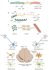
Figure 1
Figure 1
α-syn structure and pathological hallmarks of PD: (A) Schematic representation of α-syn. α-syn is divided into N-terminal, non-amyloid-beta component (NAC), and C-terminal; three domains highlighted in green, yellow, and red, respectively. Several familial PD-related mutations and post-translational modification sites are denoted. (B) Structure of α-syn monomer. (C) α-syn equilibrium. α-syn monomer can aggregate into oligomer or fibril. (D) Pathological hallmarks of PD. The pathological hallmarks of PD include progress loss of dopaminergic (DA) neurons in the substantia nigra pars compacta (SNpc), misfolded α-syn aggregates and neurites known as Lewy bodies (LBs) and Lewy neurites (LNs), and glial activation. α-syn could transfer to and activate microglia and astrocyte, which in turn release pro-/anti-inflammatory cytokines and contribute to neurodegeneration.
Figure 2
Figure 2
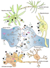
Cell-to-cell transmission of α-syn. Illustration of α-syn cell-to-cell transmission. α-syn could transfer between neurons and glia. α-syn is released via ① passive diffusion (only monomer), ② exocytosis, ③ exosomes, or ④ exophagy (grey color numbers). α-syn is taken up via ① passive diffusion (only monomer), ⑤ endocytosis, ⑥ clathrin-mediated endocytosis (CME), ⑦ receptor-mediated internalization, ⑧ micropinocytosis, ⑨ phagocytosis, or ⑩ lipid raft (black color numbers). The receptors involved in α-syn internalization include lymphocyte-activation gene 3 (LAG3), α3-subunit of Na+/K+-ATPase (α3-NKA), and the gap junction protein connexin-32 (Cx32) in neuron; Toll-like receptors 2 and 4 (TLR2 and TLR4), the scavenger receptor CD36, integrin CD11b, and the Fcγ receptors (FcγR) in microglia; and Cx32 in oligodendrocyte. In addition, α-syn can directly cell-to-cell transfer by ⑪ tunneling nanotubes (TNTs) (white color numbers). Internalized α-syn are degraded via the ubiquitin-proteasome system (UPS), autophagy, and chaperone-mediated autophagy (CMA) pathways.
Figure 3
Figure 3
Neuron-glia crosstalk in regulating the pathogenesis of α-syn in PD. Neuronal released α-syn could transfer to and activate microglia and astrocyte, activated glia in turn perform neuroprotective or neurotoxic feedback to DA neuron. Bottom left: In microglia, uptake of α-syn triggers various signaling cascades. Upon α-syn binding to heterodimer TLR1/2 receptor, it interacts with adaptor molecule MyD88 and initiates sequential IRAK/TRAF6/TAK1 activation events, then leads to MAPK activation and nuclear translocation of NF-κB, JNK, and p38, which promote the transcription of pro-inflammatory cytokines including TNF-α, IL-1β, IL-12, and inducible nitric oxide synthase (iNOS). TLR4 mediates α-syn-induced phagocytic activity, pro-inflammatory cytokine release, and ROS production. TLR4-induced NF-κB signaling upregulates the expression of p62/SQSTM1 and promote autophagy. Nod-like receptor protein 3 (NLRP3) inflammasome is involved in the inflammatory response. DJ-1 regulates α-syn phagocytosis, inflammatory responses, and degradation in microglia. LRRK2 positively regulates NF-κB inflammation pathway. Nrf2 was also reported to be involved in α-syn-induced microglia activation. Bottom right: In astrocyte, α-syn-induced pro-inflammatory cytokines, ROS production, JNK and p38 activation, and NF-κB nuclear translocation all depend on TLR4. Monomeric and oligomeric α-syn induce a Ca2+ flux via opening of connexin 43 (Cx43) hemichannels and pannexin-1 (Panx1) channels. α-syn also could triggered ER stress through PERK/eIF2α signaling pathway and CHOP-mediated apoptosis pathway. Golgi fragmentation was also observed. Nrf2 plays a neuroprotective role via reducing oxidative stress. Top right: Activated glia may perform neuroprotective roles via providing trophic factors including brain-derived neurotrophic factor (BDNF), glial-derived neurotrophic factor (GDNF), and nerve growth factor (NGF). When glia are activated, the concentration of these trophic factors decreased. Astrocyte-derived factors Wnt1 could maintain DA neuron integrity via activating Frizzled (Fzd) receptor and β-catenin signaling in DA neuron, functioning as a neuroprotective pathway. The Kir6.1/K-ATP channel on astrocytes could protect against DA neuron degeneration via promoting mitophagy, which resulted in a decrease in mitochondria accumulation, ROS production, and neuroinflammation. Top left: Activated glia may perform neurotoxic roles via production of pro-inflammatory cytokines or ROS. The pro-inflammatory cytokines TNF-α may exert a more direct effect through binding to the tumor necrosis factor receptor (TNFR) expressed by DA neurons, the intracellular death domain of TNFR1 binds to the adaptor molecules TNFR1-associated protein with a death domain (TRADD) and FAS-associated protein with a death domain (FADD), then activate the apoptosis effector caspases. Pro-inflammatory cytokines can also induce glial NO and ROS production, both of which are deleterious to DA neurons. Activated microglia also lead to severe DA neuronal degeneration by promoting phagocytic exhaustion and upregulating chemotactic molecules to selectively recruit peripheral immune cells. Oligomeric α-syn induces excessive astrocytic glutamate release, which leads to further the activation of neuronal extrasynaptic NMDA receptors (eNMDARs). Meanwhile, oligomeric α-syn also directly activates eNMDAR. The aberrant eNMDAR activity finally contributes to synaptic damage and loss.
Figure 4
Figure 4
Glial activation in regulating DA neuron ferroptosis. Iron binding could enhance α-syn aggregation by generating conformational changes in neuron. Many genes have been involved in regulating ferroptosis. NEDD4L inhibits intracellular iron accumulation and ferroptosis through binding and degrades lactotransferrin (LTF). Glutathione peroxidase 4 (GPX4) suppresses lipid peroxidation and this requires GSH. The glutamine secreted from neurons can be converted into glutamate and increase extracellular glutamate content, which in turn inhibits XcT function and results in ferroptosis. All three types of glia could regulate DA neuron ferroptosis, in either promoting or inhibiting ways. Iron accumulation induces microglia activation, activated microglia promote pro-inflammatory factors release such as TNF-α, IL-6, IL-1β, and ROS production, all of which contributes to DA neuronal death. Inflammatory cytokines produced by active microglia and astrocytes could upregulate Divalent metal transporter 1 (DMT1) and downregulate ferroportin1 (FPN1), resulting in iron accumulation in neurons. Astrocytes can help to prevent iron overload in neurons, disruptions in astrocyte-neuron connections and insufficient Nrf2 activation may result in ferroptosis in neurons. Astrocytes transmit extra GSH to the neuronal region, replenishing the antioxidant dysfunction in neurons during ferroptosis. Active astrocytes also release BDNF and GDNF to inhibited iron uptake by reducing DMT1 expression. Astrocytes and oligodendrocytes regulate glutamate levels in the synaptic cleft by regulating GSH production and suppressing neuronal XcT. Oligodendrocytes secrete FTH1 into the extracellular space to protect neurons from oxidative injury.

References

    1. Parkinson J. An essay on the shaking palsy, 1817. J. Neuropsychiatry Clin. Neurosci. 2002;14:223–236. doi: 10.1176/jnp.14.2.223. - DOI - PubMed
    1. Marras C., Beck J.C., Bower J.H., Roberts E., Ritz B., Ross G.W., Abbott R.D., Savica R., Van Den Eeden S.K., Willis A.W., et al. Prevalence of Parkinson’s disease across North America. npj Park. Dis. 2018;4:21. doi: 10.1038/s41531-018-0058-0. - DOI - PMC - PubMed
    1. de Rijk M.C., Breteler M.M., Graveland G.A., Ott A., Grobbee D.E., van der Meche F.G., Hofman A. Prevalence of Parkinson’s disease in the elderly: The Rotterdam Study. Neurology. 1995;45:2143–2146. doi: 10.1212/WNL.45.12.2143. - DOI - PubMed
    1. Barbeau A. L-dopa therapy in Parkinson’s disease: A critical review of nine years’ experience. Can. Med. Assoc. J. 1969;101:59–68. - PMC - PubMed
    1. Postuma R.B., Berg D., Stern M., Poewe W., Olanow C.W., Oertel W., Obeso J., Marek K., Litvan I., Lang A.E., et al. MDS clinical diagnostic criteria for Parkinson’s disease. Mov. Disord. 2015;30:1591–1601. doi: 10.1002/mds.26424. - DOI - PubMed

MeSH terms

Substances