. 2022 Nov 25;23(23):14769.
doi: 10.3390/ijms232314769.

Understanding In Vitro Pathways to Drug Discovery for TDP-43 Proteinopathies

Affiliations

Understanding In Vitro Pathways to Drug Discovery for TDP-43 Proteinopathies

Hei W A Cheng et al. Int J Mol Sci. .

Abstract

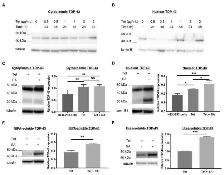
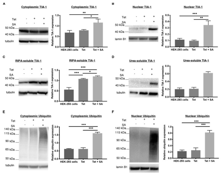
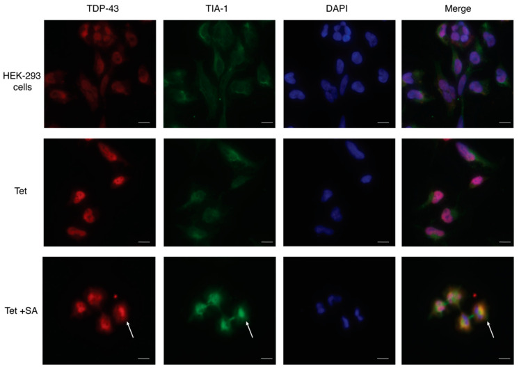
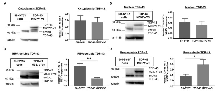
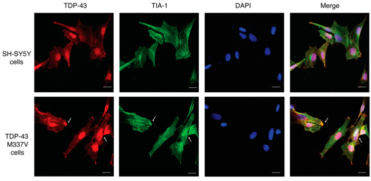
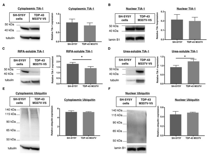
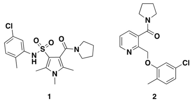
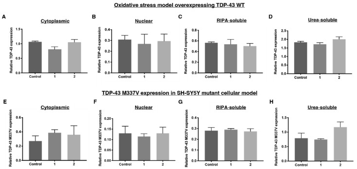
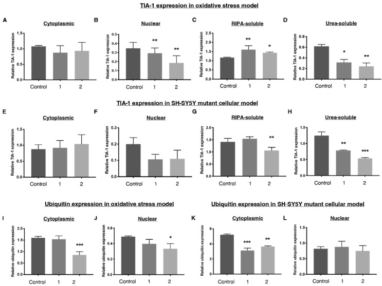
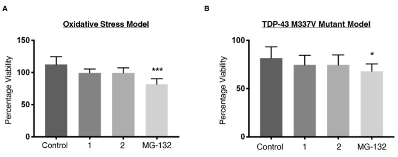
The use of cellular models is a common means to investigate the potency of therapeutics in pre-clinical drug discovery. However, there is currently no consensus on which model most accurately replicates key aspects of amyotrophic lateral sclerosis (ALS) and frontotemporal dementia (FTD) pathology, such as accumulation of insoluble, cytoplasmic transactive response DNA-binding protein (TDP-43) and the formation of insoluble stress granules. Given this, we characterised two TDP-43 proteinopathy cellular models that were based on different aetiologies of disease. The first was a sodium arsenite-induced chronic oxidative stress model and the second expressed a disease-relevant TDP-43 mutation (TDP-43 M337V). The sodium arsenite model displayed most aspects of TDP-43, stress granule and ubiquitin pathology seen in human ALS/FTD donor tissue, whereas the mutant cell line only modelled some aspects. When these two cellular models were exposed to small molecule chemical probes, different effects were observed across the two models. For example, a previously disclosed sulfonamide compound decreased cytoplasmic TDP-43 and increased soluble levels of stress granule marker TIA-1 in the cellular stress model without impacting these levels in the mutant cell line. This study highlights the challenges of using cellular models in lead development during drug discovery for ALS and FTD and reinforces the need to perform assessments of novel therapeutics across a variety of cell lines and aetiological models.

Keywords: TDP-43; amyotrophic lateral sclerosis; frontotemporal dementia; proteinopathy; stress granules.


Conflict of interest statement

The authors declare no conflict of interest.

Figures

Figure 1
Figure 1
Comparing TDP-43 expression levels in human embryonic kidney (HEK-293) cells stably transfected with tetracycline (Tet)–inducible WT TDP-43 using Western blots. Cells were treated with varied concentrations of Tet (μg/mL) for different time periods. (A,B) The Western blot shows overexpression of TDP-43 in the cytoplasm and nucleus of transfected HEK-293 cells treated with 2 μg/mL of Tet for 48 h. (C,D) The quantified expression of TDP-43 in the cytoplasm and nucleus after treatment with Tet (2 μg/mL, 48 h) with and without sodium arsenite (SA, 15 μM, 18 h), an oxidative stressor, to encourage TDP-43 translocation and formation of stress granules. (E,F) RIPA- and urea-soluble fractions of transfected HEK-293 cells were stained for TDP-43. TDP-43 is normalised to α-tubulin for the cytoplasmic, RIPA-soluble and urea-soluble fractions and lamin B1 for the nuclear fraction. Data represent mean ± SD, n ≥ 3, and significant differences between means were examined with a one-way ANOVA with Tukey’s multiple comparison test. * p < 0.05, ** p < 0.01, *** p < 0.001, ns: not significant.
Figure 2
Figure 2
Comparing TIA-1 and ubiquitin expression levels in stably transfected HEK-293 cells. The expression level of the stress granule marker, TIA-1 in stably transfected HEK-293 cells with and without tetracycline (Tet, 2 μg/mL, 48 h) and sodium arsenite (SA, 15 μM, 18 h) was measured using Western blots. (A,B) The quantified, relative expression levels of TIA-1 in the cytoplasm and nucleus. (C,D) RIPA- and urea-soluble TIA-1 expression. The Western blots and corresponding histograms showing ubiquitin expression levels in the (E,F) cytoplasm and nucleus of HEK-293 cells. TIA-1 and ubiquitin are normalised to α-tubulin for the cytoplasmic, RIPA-soluble and urea-soluble fraction and lamin B1 for the nuclear fraction. Data represent mean ± SD, n ≥ 3, and significant differences between means were examined with a one-way ANOVA with Tukey’s multiple comparison test. * p < 0.05, ** p < 0.01, *** p < 0.001.
Figure 3
Figure 3
Immunofluorescence imaging of TDP-43 and TIA-1 in stably transfected HEK-293 cells. HEK-293 cells transfected with tetracycline (Tet)–inducible WT TDP-43 (2 ug/mL, 48 h) resulting in overexpression of TDP-43 (red), especially in the nucleus, but low levels of TIA-1. Sodium arsenite (15 μM, 18 h) treated cells displayed mislocalisation of TDP-43 which colocalised with upregulated TIA-1 (green) in the cytoplasm (white arrow). The nuclear stain DAPI (blue) was used to indicate the nucleus. Scale bar = 10 μM.
Figure 4
Figure 4
Comparing TDP-43 distribution in human neuroblastoma SH-SY5Y cells stably transfected with TDP-43 M337V mutant (V5-tagged) compared to WT SH-SY5Y cells using Western blots. (A,B) Cytoplasmic and nuclear TDP-43 expression of SH-SY5Y WT cells compared to transfected SH-SY5Y TDP-43 M337V. (C,D) Relative RIPA-soluble and urea-soluble expression levels of TDP-43 WT in SH-SY5Y cells compared to TDP-43 M337V mutant. Data represent mean ± SD, n ≥ 3, statistical analysis using parametric, unpaired t-test. * p < 0.05, *** p < 0.001.
Figure 5
Figure 5
Immunofluorescence imaging of wild–type human neuroblastoma SH-SY5Y cells compared to SH-SY5Y cells expressing the ALS–linked mutation TDP-43 M337V. Representative images of SH-SY5Y cells and M337V TDP-43 SH-SY5Y cells showing similar expression levels and distribution of TDP-43 (red), stress granule marker, and TIA-1 (green). DAPI (blue) was used to indicate the nucleus. The arrow shows the colocalisation of TDP-43 and stress granule aggregates. Scale bar = 10 μM.
Figure 6
Figure 6
Comparing TIA-1 distribution in wild-type human neuroblastoma SH-SY5Y cells and SH-SY5Y cells stably transfected with TDP-43 M337V mutant (V5-tagged) using Western blots. (A,B) Cytoplasmic and nuclear TIA-1 expression in SH-SY5Y WT cells compared to transfected SH-SY5Y TDP-43 M337V. The expression of TIA-1 did not significantly change after TDP-43 M337V transfection (C,D) The solubility of TIA-1 in SH-SY5Y cells compared to TDP-43 M337V mutant. Ubiquitin expression in the (E) cytoplasm and (F) nucleus. The presence of TDP-43 M337V transfection did not change the level of ubiquitin throughout the cell. Data represent mean ± SD, n ≥ 3, statistical analysis using parametric, unpaired t-test. * p < 0.05.
Figure 7
Figure 7
Chemical structures of 1 and 2.
Figure 8
Figure 8
Relative TDP-43 WT expression levels after treatment with 1 and 2 as determined by Western blots. (A) Relative TDP-43 WT expression in the cytoplasm, (B) nucleus, (C) RIPA-soluble fraction and (D) urea-soluble fraction after the oxidative stress HEK-293 cellular model, featuring TDP-43 overexpressing HEK-293 cells treated with tetracycline (2 ug/mL, 48 h) and sodium arsenite (15 μM, 18 h), which was exposed to 1 and 2. Relative TDP-43 M337V-V5 expression levels in the (E) cytoplasm, (F) nucleus, (G) RIPA-soluble fraction and (H) urea-soluble fraction from SH-SY5Y TDP-43 M337V mutant cellular model after treatment of 1 and 2. Both cell lines were treated with 1 and 2 (300 nM) for 18 h. Densitometry of TDP-43 WT and M337V are normalised to α-tubulin for the cytoplasmic, RIPA-soluble and urea-soluble fraction and lamin B1 for the nuclear fraction. Data represent mean ± SD, n ≥ 3. Significant differences were indexed using a one-way ANOVA with Dunnett’s post hoc test.
Figure 9
Figure 9
Relative distribution of TIA-1, a stress granule marker and ubiquitin in both cellular models as determined by Western blots. Relative distribution of TIA-1 expression in the (A) cytoplasm, (B) nucleus, (C) RIPA-soluble fraction and (D) urea-soluble fraction of the oxidative stress model, featuring HEK-293 cells overexpressing TDP-43 WT, which were treated with tetracycline (2 ug/mL, 48 h) and sodium arsenite (15 μM, 18 h). Relative distribution of TIA-1 in the SH-SY5Y TDP-43 M337V cells in the (E) cytoplasm, (F) nucleus, (G) RIPA-soluble fraction and (H) urea-soluble fraction. (I,J) Ubiquitin expression in the cytoplasm and nucleus of the oxidative stress model overexpressing TDP-43 WT. (K,L) Ubiquitin expression in the cytoplasm and nucleus of the SH-SY5Y TDP-43 M337V model. Both cell lines were treated with 1 and 2 (300 nM) for 18 h. Densitometry of TIA-1 and ubiquitin are normalised to α-tubulin for the cytoplasmic, RIPA-soluble and urea-soluble fraction and lamin B1 for the nuclear fraction. Data represent mean ± SD, n ≥ 3. Significant differences between means were assessed by a one-way ANOVA with Dunnett’s post hoc test. * p < 0.05, ** p < 0.01, *** p < 0.001.
Figure 10
Figure 10
CellTiter-Blue® cell viability assay. (A) Results were calculated as a percentage of HEK-293 cells treated with only tetracycline and not sodium arsenite. (B) The percentage viability is expressed as a percentage of untreated wild-type SH-SY5Y cells. Both cell lines were treated with 1, 2 (300 nM) or MG-132 (5 μM) for 18 h. Data represent mean ± SD, n≥ 3. Significant differences were tested using one-way ANOVA with Dunnett’s post-hoc test, * p < 0.05, *** p < 0.001.

Similar articles

Cited by

  • Molecular Mechanisms of Dementia.
    Cantone M. Cantone M. Int J Mol Sci. 2023 Aug 22;24(17):13027. doi: 10.3390/ijms241713027. Int J Mol Sci. 2023. PMID: 37685834 Free PMC article.

References

    1. Neumann M., Sampathu D.M., Kwong L.K., Truax A.C., Micsenyi M.C., Chou T.T., Bruce J., Schuck T., Grossman M., Clark C.M. Ubiquitinated TDP-43 in frontotemporal lobar degeneration and amyotrophic lateral sclerosis. Science. 2006;314:130–133. doi: 10.1126/science.1134108. - DOI - PubMed
    1. Mackenzie I.R., Bigio E.H., Ince P.G., Geser F., Neumann M., Cairns N.J., Kwong L.K., Forman M.S., Ravits J., Stewart H. Pathological TDP-43 distinguishes sporadic amyotrophic lateral sclerosis from amyotrophic lateral sclerosis with SOD1 mutations. Ann. Neurol. 2007;61:427–434. doi: 10.1002/ana.21147. - DOI - PubMed
    1. Hasegawa M., Arai T., Nonaka T., Kametani F., Yoshida M., Hashizume Y., Beach T.G., Buratti E., Baralle F., Morita M. Phosphorylated TDP-43 in frontotemporal lobar degeneration and amyotrophic lateral sclerosis. Ann. Neurol. 2008;64:60–70. doi: 10.1002/ana.21425. - DOI - PMC - PubMed
    1. Jain S., Wheeler J.R., Walters R.W., Agrawal A., Barsic A., Parker R. ATPase-modulated stress granules contain a diverse proteome and substructure. Cell. 2016;164:487–498. doi: 10.1016/j.cell.2015.12.038. - DOI - PMC - PubMed
    1. McGurk L., Gomes E., Guo L., Mojsilovic-Petrovic J., Tran V., Kalb R.G., Shorter J., Bonini N.M. Poly (ADP-ribose) prevents pathological phase separation of TDP-43 by promoting liquid demixing and stress granule localization. Mol. Cell. 2018;71:703–717. doi: 10.1016/j.molcel.2018.07.002. - DOI - PMC - PubMed

MeSH terms

Substances