

Innate Immune System Activation, Inflammation and Corneal Wound Healing
- PMID: 36499260
- PMCID: PMC9740891
- DOI: 10.3390/ijms232314933
Innate Immune System Activation, Inflammation and Corneal Wound Healing
Abstract
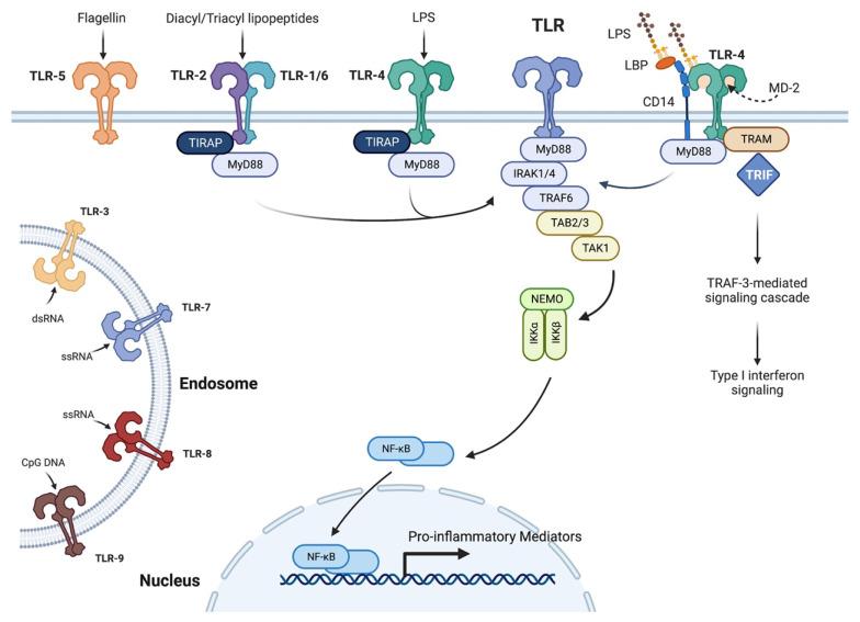
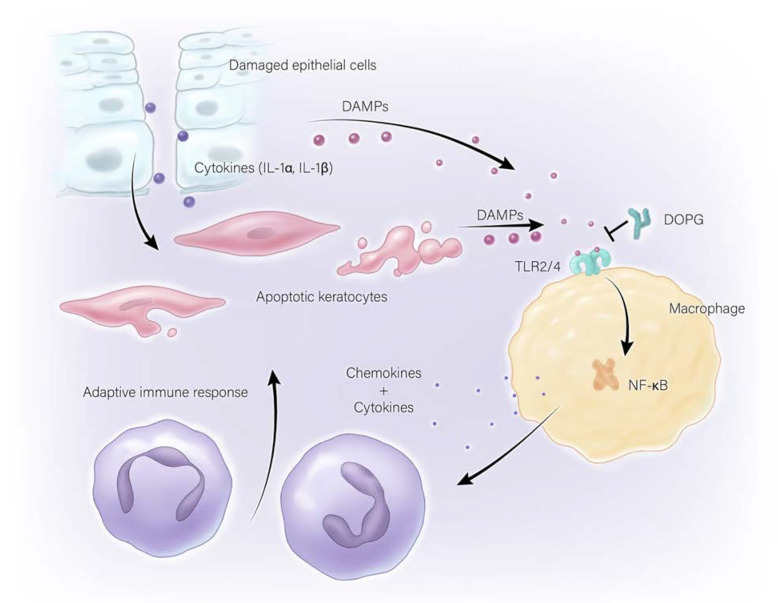
Corneal wounds resulting from injury, surgeries, or other intrusions not only cause pain, but also can predispose an individual to infection. While some inflammation may be beneficial to protect against microbial infection of wounds, the inflammatory process, if excessive, may delay corneal wound healing. An examination of the literature on the effect of inflammation on corneal wound healing suggests that manipulations that result in reductions in severe or chronic inflammation lead to better outcomes in terms of corneal clarity, thickness, and healing. However, some acute inflammation is necessary to allow efficient bacterial and fungal clearance and prevent corneal infection. This inflammation can be triggered by microbial components that activate the innate immune system through toll-like receptor (TLR) pathways. In particular, TLR2 and TLR4 activation leads to pro-inflammatory nuclear factor kappa-light-chain-enhancer of activated B cells (NFκB) activation. Similarly, endogenous molecules released from disrupted cells, known as damage-associated molecular patterns (DAMPs), can also activate TLR2, TLR4 and NFκB, with the resultant inflammation worsening the outcome of corneal wound healing. In sterile keratitis without infection, inflammation can occur though TLRs to impact corneal wound healing and reduce corneal transparency. This review demonstrates the need for acute inflammation to prevent pathogenic infiltration, while supporting the idea that a reduction in chronic and/or excessive inflammation will allow for improved wound healing.
Keywords: cornea; healing; inflammation; innate immune system; phosphatidylglycerol; toll-like receptors; wound.
Conflict of interest statement
WBB is an inventor of an Augusta University patent for the use of dioleoyl-phosphatidylglycerol to accelerate corneal wound healing. All other authors report no conflict of interest. The funders had no role in the design of the study; in the collection, analyses, or interpretation of data; in the writing of the manuscript; or in the decision to publish the results.
Figures
Similar articles
-
Toll-like receptor 2 siRNA suppresses corneal inflammation and attenuates Aspergillus fumigatus keratitis in rats.Immunol Cell Biol. 2012 Mar;90(3):352-7. doi: 10.1038/icb.2011.49. Epub 2011 Jun 7. Immunol Cell Biol. 2012. PMID: 21647173
-
Modulation of immune signaling, bacterial clearance, and corneal integrity by toll-like receptors during streptococcus pneumoniae keratitis.Curr Eye Res. 2013 Oct;38(10):1036-48. doi: 10.3109/02713683.2013.804094. Epub 2013 Jul 10. Curr Eye Res. 2013. PMID: 23841825 Free PMC article.
-
Small leucine-rich repeat proteoglycans in corneal inflammation and wound healing.Exp Eye Res. 2016 Oct;151:142-9. doi: 10.1016/j.exer.2016.08.015. Epub 2016 Aug 26. Exp Eye Res. 2016. PMID: 27569372 Free PMC article. Review.
-
Thymosin beta 4: A potential novel adjunct treatment for bacterial keratitis.Int Immunopharmacol. 2023 May;118:109953. doi: 10.1016/j.intimp.2023.109953. Epub 2023 Apr 3. Int Immunopharmacol. 2023. PMID: 37018981 Free PMC article.
-
Role of aquaporins in corneal healing post chemical injury.Exp Eye Res. 2023 Mar;228:109390. doi: 10.1016/j.exer.2023.109390. Epub 2023 Jan 22. Exp Eye Res. 2023. PMID: 36696947 Free PMC article. Review.
Cited by
-
Consumption of Limosilactobacillus fermentum Inhibits Corneal Damage and Inflammation in Dry Eye Disease Mouse Model through Regulating the Gut Microbiome.Int J Mol Sci. 2024 Mar 20;25(6):3528. doi: 10.3390/ijms25063528. Int J Mol Sci. 2024. PMID: 38542500 Free PMC article.
-
Topical losartan ophthalmic drops - a review of corneal wound healing and topical losartan for managing corneal haze and potential future indications.Graefes Arch Clin Exp Ophthalmol. 2025 Apr;263(4):925-934. doi: 10.1007/s00417-024-06710-8. Epub 2024 Dec 12. Graefes Arch Clin Exp Ophthalmol. 2025. PMID: 39665995
-
The Role of Macrophage Migration Inhibitory Factor (MIF) and D-Dopachrome Tautomerase (D-DT/MIF-2) in Infections: A Clinical Perspective.Biomedicines. 2023 Dec 19;12(1):2. doi: 10.3390/biomedicines12010002. Biomedicines. 2023. PMID: 38275363 Free PMC article. Review.
-
Recognition of Mycobacterium tuberculosis by macrophage Toll-like receptor and its role in autophagy.Inflamm Res. 2024 May;73(5):753-770. doi: 10.1007/s00011-024-01864-x. Epub 2024 Apr 2. Inflamm Res. 2024. PMID: 38563966 Review.
-
Aquaporins in the Cornea.Int J Mol Sci. 2024 Mar 28;25(7):3748. doi: 10.3390/ijms25073748. Int J Mol Sci. 2024. PMID: 38612559 Free PMC article. Review.
References
-
- Mescher A.L. Junqueira’s Basic Histology: Text and Atlas. 15th ed. McGraw-Hill Education; New York, NY, USA: 2018. The Eye & Ear: Special Sense Organs.
-
- Lee T.N. The ins and outs of corneal wound healing. [(accessed on 23 August 2022)];Rev. Optom. 2016 Available online: https://www.reviewofoptometry.com/article/the-ins-and-outs-of-corneal-wo....
-
- Alshamahi E.Y.A., Al Nahary A.A., Al Shamahy H.A., Al Magrami R.T.F., Alhowthi M.A. Epidemiology and aetiological diagnosis of corneal ulceration in Sana’a City, Yemen. World J. Ophthalmol. Vis. Res. 2019;2 doi: 10.33552/WJOVR.2019.02.000550. - DOI
Publication types
MeSH terms
Substances
Grants and funding
LinkOut - more resources
Full Text Sources
Medical
