. 2022 Nov 29;23(23):14947.
doi: 10.3390/ijms232314947.

A New Organotypic 3D Slice Culture of Mouse Meibomian Glands Reveals Impact of Melanocortins

Affiliations

A New Organotypic 3D Slice Culture of Mouse Meibomian Glands Reveals Impact of Melanocortins

Ingrid Zahn et al. Int J Mol Sci. .

Abstract

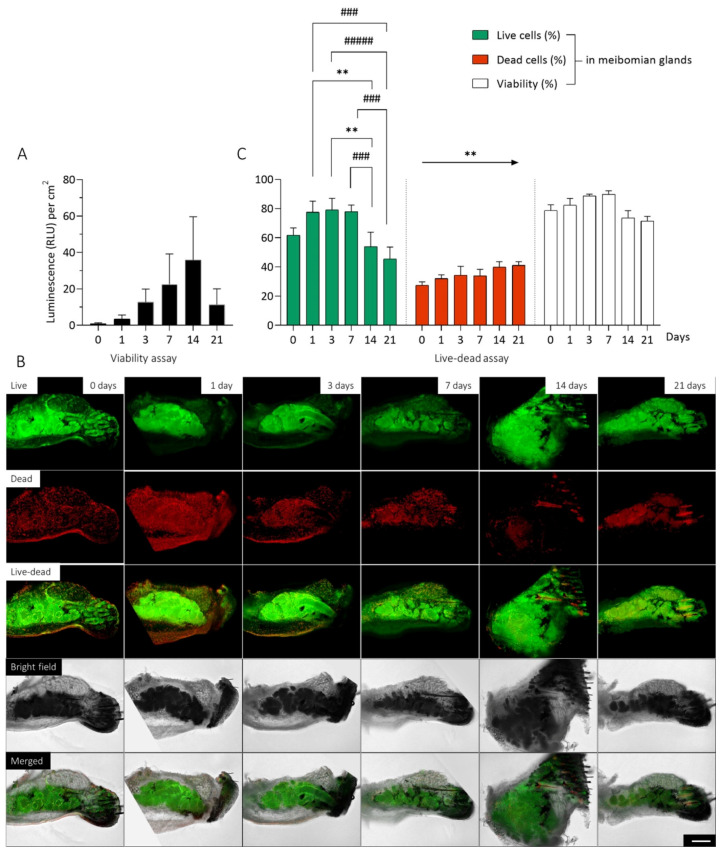
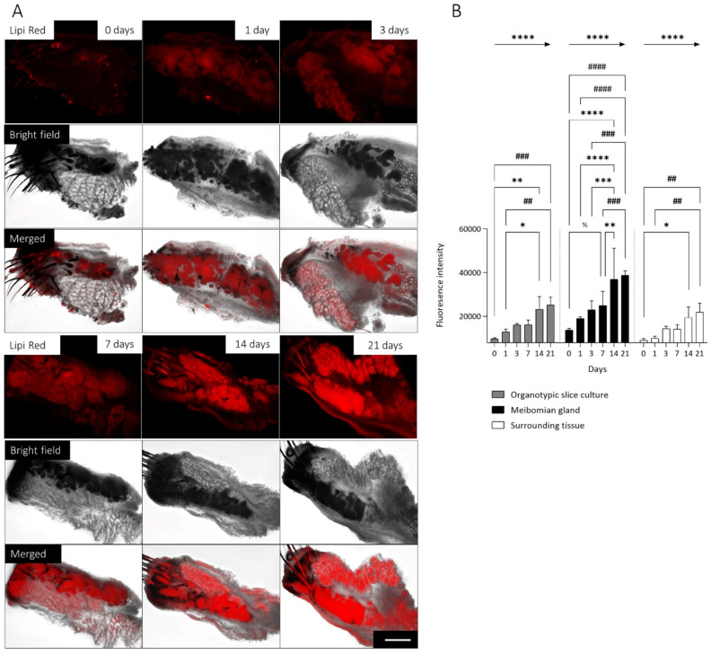
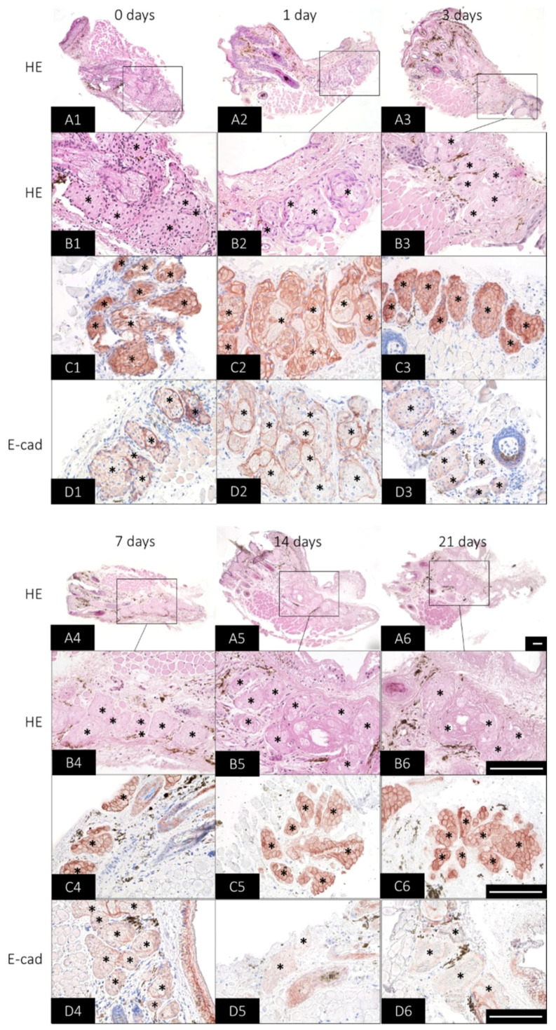
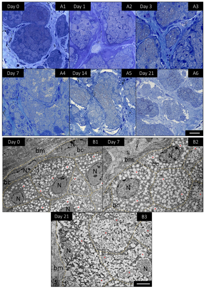
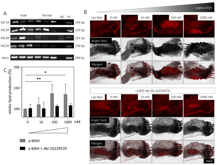
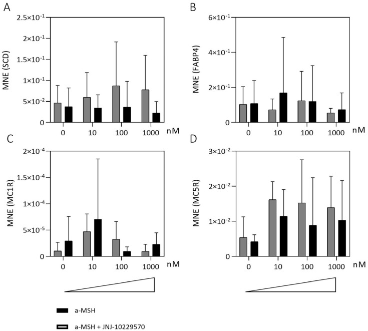
The meibomian glands (MGs) within the eyelids produce a lipid-rich secretion that forms the superficial layer of the tear film. Meibomian gland dysfunction (MGD) results in excessive evaporation of the tear film, which is the leading cause of dry eye disease (DED). To develop a research model similar to the physiological situation of MGs, we established a new 3D organotypic slice culture (OSC) of mouse MGs (mMGs) and investigated the effects of melanocortins on exocrine secretion. Tissue viability, lipid production and morphological changes were analyzed during a 21-day cultivation period. Subsequently, the effects on lipid production and gene expression were examined after stimulation with a melanocortin receptor (MCR) agonist, α-melanocyte-stimulating hormone (α-MSH), and/or an MCR antagonist, JNJ-10229570. The cultivation of mMGs OSCs was possible without impairment for at least seven days. Stimulation with the MCR agonists induced lipid production in a dose-dependent manner, whereas this effect was tapered with the simultaneous incubation of the MCR antagonist. The new 3D OSC model is a promising approach to study the (patho-) physiological properties of MG/MGD while reducing animal studies. Therefore, it may accelerate the search for new treatments for MGD/DED and lead to new insights, such as that melanocortins likely stimulate meibum production.

Keywords: 3D cell culture model; dry eye disease; meibomian gland; meibomian gland dysfunction; melanocortin receptor; organotypic slice culture; vibratome; α-MSH.


Conflict of interest statement

The authors declare that they have no conflict of interest.

Figures

Figure 1
Figure 1
Viability of organotypic slice cultures (OSC) of mouse meibomian glands (mMG) over 21 days. The viability of mMG OSCs were investigated over 21 days. No decrease in viability was observed within the first 14 days of cultivation (A). Representative pictures of OSCs after live-dead staining illustrates the location and distribution of the live (green) and dead (red) cells. Bright field imaging shows the position of the mMGs within the OSC (B). The evaluation of live-dead images based on the proportion of live and dead cells and the viability of mMG during the cultivation period (C). Most of the live cells are located in the meibomian glands, while the dead cells are homogeneously distributed throughout the tissue. Data are n = 9 (A) and 4 (B,C), mean ± SEM. Significant changes compared to day 14 are assigned as */compared to day 21 are assigned as #. An arrow indicates a linear trend test. ** p ≤ 0.01, ### p ≤ 0.001, ##### p ≤ 0.0001 (C). Scale bar: 200 µm (B).
Figure 2
Figure 2
Lipid production in organotypic slice cultures (OSC) of mouse meibomian glands (mMG) over 21 days. Representative pictures of lipid droplets visualized by Lipi Red staining of mMG OSCs for 21 days. Bright field imaging shows the position of the mMGs within the OSC. Most lipid droplets are located in the mMGs (A). During the cultivation period, the fluorescence intensity increases significant linearly in the whole OSC, for the mMG as well as the surrounding tissue (B). Significant changes compared to day 7 are assigned as %/day 14 are assigned as */day 21 are assigned as #. Arrows indicate a linear trend test. Data are n = 4, mean ± SD, */% p ≤ 0.05, **/## p ≤ 0.01, ***/### p ≤ 0.001, ****/#### p ≤ 0.0001 (B). Scale bar: 200 µm (A).
Figure 3
Figure 3
Histological changes of organotypic slice cultures (OSC) of mouse meibomian glands (mMG) over 21 days. Representative pictures of mMG OSC morphology visualized by hematoxylin and eosin (HE), cytokeratin 14 (CK14) and E-cadherin (E-cad)-staining for 21 days. HE at 100× magnification serves as an overview staining for the OSC (A1A6). The 400× magnification shows that as the cultivation time progresses, fewer nuclei can be stained in the mMG acini (*) (B1B6). CK14 antibody reaction is maintained in the meibomian gland acini (*) over the culture period (C1C6), whereas E-cad immunoreaction decreases at 14 and 21 days (D1D6). Data are n = 3, scale bar = 100 µm (A1D6).
Figure 4
Figure 4
Detailed morphological changes of mouse meibomian gland (mMG) acini in organotypic slice cultures (OSC) over 21 days. Representative pictures of toluidine blue stained semi-thin sections of mMG OSCs for 21 days (A1A6). Ultrathin sections analyzed by transmission electron microscopy revealed that basal cells (bc) located next to the basal membrane (bm) enlarge and undergo lipogenesis with increasing cultivation time. Thereby, the bc accumulate more lipids (*) and the nucleus (N) becomes increasingly pyknotic (day 0, day 7 and day 21) (B1B3). Data are n = 3, Scale bar = 25 µm (A1A6), 5 µm (B1B3).
Figure 5
Figure 5
Influence of a melanocortin receptor (MCR) agonist (α-MSH) and MCR antagonist (JNJ-10229570) on lipid production in mouse meibomian gland (mMG) organotypic slice culture (OSC). RT-PCR detection of MC1R, MC3R, MC4R and MC5R mRNA in male and female mMG from three different mice (A). Representative pictures of lipid droplets visualized by Lipi Red staining of OSCs treated with medium containing up to 1000 nM α-MSH with/without 1000 nM JNJ-10229570 for 1 day (B). Lipid production increases significantly with the addition of 100 nM and 1000 nM α-MSH, whereas this effect is absent when JNJ-10229570 is added (C). Data are n = 4, mean ± SD, NC = negative control, PC = positive control (mouse brain). Scale bar: 200 µm * p ≤ 0.05, ** p ≤ 0.01.
Figure 6
Figure 6
Influence of a melanocortin receptor (MCR) agonist (α-MSH) and MCR antagonist (JNJ-10229570) on gene expression levels in mouse meibomian gland (mMG) organotypic slice culture (OSC). Gene expression levels of OSCs treated with medium containing up to 1000 nM α-MSH with/without 1000 nM JNJ-10229570 for 1 day. Mean normalized expression levels (MNE) of Stearoyl-CoA desaturase (SCD) (A), fatty acid-binding protein 4 (FABP4) (B), melanocortin receptor 1 (MC1R) (C) and melanocortin receptor 5 (MC5R) (D). Stimulation with α-MSH tends to increase gene expression levels of SCD and MC5R but not of MC1R and FABP4. Data are n = 6 (A,B) n = 3 (C,D), mean ± SEM.

Similar articles

Cited by

References

    1. Jester J.V., Nicolaides N., Smith R.E. Meibomian gland studies: Histologic and ultrastructural investigations. Investig. Ophthalmol. Vis. Sci. 1981;20:537–547. - PubMed
    1. Kozak I., Bron A.J., Kucharova K., Kluchova D., Marsala M., Heichel C.W., Tiffany J.M. Morphologic and volumetric studies of the meibomian glands in elderly human eyelids. Cornea. 2007;26:610–614. doi: 10.1097/ICO.0b013e318041f0d2. - DOI - PubMed
    1. Greiner J.V., Glonek T., Korb D.R., Whalen A.C., Hebert E., Hearn S.L., Esway J.E., Leahy C.D. Volume of the human and rabbit meibomian gland system. Adv. Exp. Med. Biol. 1998;438:339–343. doi: 10.1007/978-1-4615-5359-5_48. - DOI - PubMed
    1. Knop E., Knop N., Millar T., Obata H., Sullivan D.A. The international workshop on meibomian gland dysfunction: Report of the subcommittee on anatomy, physiology, and pathophysiology of the meibomian gland. Investig. Ophthalmol. Vis. Sci. 2011;52:1938–1978. doi: 10.1167/iovs.10-6997c. - DOI - PMC - PubMed
    1. Olami Y., Zajicek G., Cogan M., Gnessin H., Pe’er J. Turnover and migration of meibomian gland cells in rats’ eyelids. Ophthalmic Res. 2001;33:170–175. doi: 10.1159/000055665. - DOI - PubMed

LinkOut - more resources