. 2022 Nov 30;23(23):15040.
doi: 10.3390/ijms232315040.

B4GALT1 as a New Biomarker of Idiopathic Pulmonary Fibrosis

Affiliations

B4GALT1 as a New Biomarker of Idiopathic Pulmonary Fibrosis

Claudia De Vitis et al. Int J Mol Sci. .

Abstract

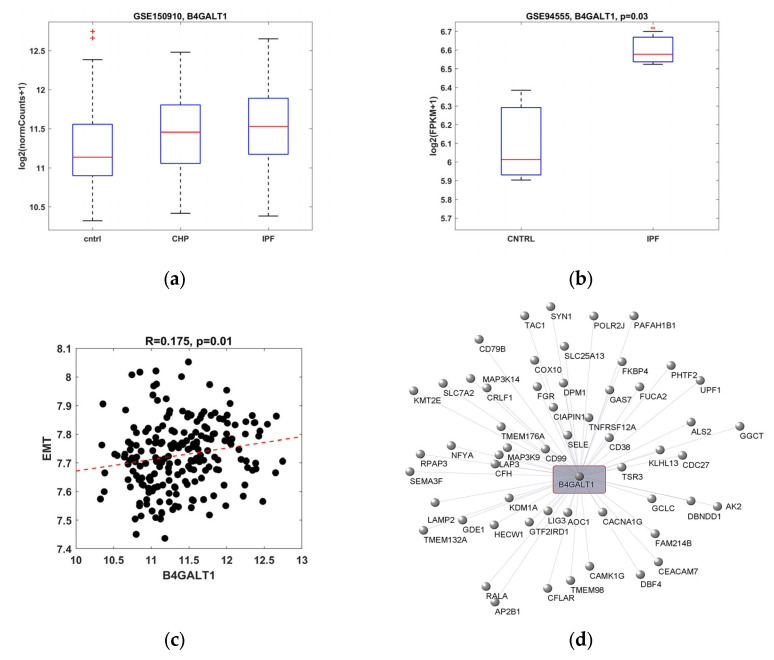
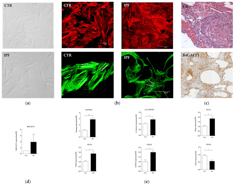
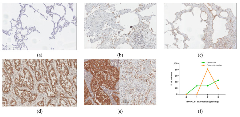
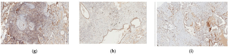
Idiopathic pulmonary fibrosis (IPF) is a disease characterized by progressive scarring of the lung that involves the pulmonary interstitium. The disease may rapidly progress, leading to respiratory failure, and the long-term survival is poor. There are no accurate biomarkers available so far. Our aim was to evaluate the expression of the B4GALT1 in patients with IPF. Analysis of B4GALT1 gene expression was performed in silico on two gene sets, retrieved from the Gene Expression Omnibus database. Expression of B4GALT1 was then evaluated, both at the mRNA and protein levels, on lung specimens obtained from lung biopsies of 4 IPF patients, on one IPF-derived human primary cell and on 11 cases of IPF associated with cancer. In silico re-analysis demonstrated that the B4GALT1 gene was overexpressed in patients and human cell cultures with IPF (p = 0.03). Network analysis demonstrated that B4GALT1 upregulation was correlated with genes belonging to the EMT pathway (p = 0.01). The overexpression of B4GALT1 was observed, both at mRNA and protein levels, in lung biopsies of our four IPF patients and in the IPF-derived human primary cell, in other fibrotic non-lung tissues, and in IPF associated with cancer. In conclusion, our results indicate that B4GALT1 is overexpressed in IPF and could represent a novel marker of this disease.

Keywords: B4GALT1; EMT; idiopathic fibrosis pulmonary; immunohistochemistry; lung cancer; lung fibrosis; mRNA.


Conflict of interest statement

There is no conflict of interest that could be perceived as prejudicing the impartiality of the research reported.

Figures

Figure 1
Figure 1
Bioinformatic analysis to evaluate B4GALT1 in silico expression. (a) Box plot representing B4GALT1 expression from RNA-seq-based transcriptomic analysis for whole lung tissues from 82 chronic hypersensitivity pneumonitis (CHP), 103 IPF, and 103 control subjects. We observed a statistical significance comparing both CHP and IPF samples to healthy subjects (p = 7 × 10⁻³, p = 4.1 × 10⁻⁵, respectively) by using the Wilcoxon rank sum test. No difference was revealed between CHP and IPF samples (p = 0.15). A Kruskal–Wallis test among three groups was also evaluated (p = 1.2 × 10⁻⁴). Data from the GEO database with access ID GSE150910. (b) Box plot representing B4GALT1 expression from scRNA-seq transcriptomic analysis of normal and IPF respiratory epithelial cells obtained from peripheral lung of controls (n = 3) and IPF patients (n = 3), respectively. Statistical significance was assessed by Student’s t-test. Data from GEO database with access ID GSE94555. (c) Scatter plot of Pearson’s correlation between B4GALT1 expression and the mean expression of HALLMARK_EPITHELIAL_MESENCHYMAL_ TRANSITION genes. Data from GEO database with access ID GSE150910. (d) Graphical network analysis obtained by Pearson’s correlation between B4GALT1 and 55 positive correlated genes involved in the EMT pathway.
Figure 2
Figure 2
In Vitro characterization of IPF primary cell line. (a) Primary culture of human normal fibroblast (upper) and IPF (lower) (×10 magnification). (b) Representative bright field images on actin of normal fibroblast and IPF primary cell cultures (×40 magnification). (c) H&E corresponding to lung biopsy of the patient with UIP from which the primary cell culture was isolated and IHC analysis (×20 magnification). (d) mRNA expression level of B4GALT1. (e) mRNA levels of stemness genes and EMT genes between normal fibroblast and IPF verified by qRT-PCR. The data are expressed as mean ± SD.* p < 0.05; ** p < 0.005.
Figure 3
Figure 3
B4GALT1 expression in lung fibrosis and lung cancer (×20 magnification). (a) An example of near-normal lung tissue is shown (B4GALT1 staining); (b) positive staining in lung cancer; (c) tumor (on the left) and UIP (on the right) of the same patient show simultaneous strong cytoplasm B4GALT1 expression; (d) strong B4GALT1 expression is observed in most fibroblastic foci in UIP both in connective tissue and alveolar epithelium; (e) and possibly in UIP fibrous reactive remodeling of lung tissue not related to IPF; (f) quantification of grading expression of B4GALT1; (g) there are B4GALT1-positive immune lymphocyte infiltrating cells in neoplastic and UIP tissue samples; and (h,i) capillary endothelium expressing B4GALT1 was observed, in the same patient, both in the immediately peritumoral connective tissue area and adjacent to the UIP pattern fibrous.
Figure 3
Figure 3
B4GALT1 expression in lung fibrosis and lung cancer (×20 magnification). (a) An example of near-normal lung tissue is shown (B4GALT1 staining); (b) positive staining in lung cancer; (c) tumor (on the left) and UIP (on the right) of the same patient show simultaneous strong cytoplasm B4GALT1 expression; (d) strong B4GALT1 expression is observed in most fibroblastic foci in UIP both in connective tissue and alveolar epithelium; (e) and possibly in UIP fibrous reactive remodeling of lung tissue not related to IPF; (f) quantification of grading expression of B4GALT1; (g) there are B4GALT1-positive immune lymphocyte infiltrating cells in neoplastic and UIP tissue samples; and (h,i) capillary endothelium expressing B4GALT1 was observed, in the same patient, both in the immediately peritumoral connective tissue area and adjacent to the UIP pattern fibrous.

References

    1. Ma H., Wu X., Li Y., Xia Y. Research Progress in the Molecular Mechanisms, Therapeutic Targets, and Drug Development of Idiopathic Pulmonary Fibrosis. Front. Pharmacol. 2022;13:963054. doi: 10.3389/fphar.2022.963054. - DOI - PMC - PubMed
    1. Ruaro B., Matucci Cerinic M., Salton F., Baratella E., Confalonieri M., Hughes M. Editorial: Pulmonary fibrosis: One manifestation, various diseases. Front. Pharmacol. 2022;13:1027332. doi: 10.3389/fphar.2022.1027332. - DOI - PMC - PubMed
    1. Calabrese F., Lunardi F., Tauro V., Pezzuto F., Fortarezza F., Vedovelli L., Faccioli E., Balestro E., Schiavon M., Esposito G., et al. RNA Sequencing of Epithelial Cell/Fibroblastic Foci Sandwich in Idiopathic Pulmonary Fibrosis: New Insights on the Signaling Pathway. Int. J. Mol. Sci. 2022;23:3323. doi: 10.3390/ijms23063323. - DOI - PMC - PubMed
    1. Confalonieri P., Volpe M.C., Jacob J., Maiocchi S., Salton F., Ruaro B., Confalonieri M., Braga L. Regeneration or Repair? The Role of Alveolar Epithelial Cells in the Pathogenesis of Idiopathic Pulmonary Fibrosis (IPF) Cells. 2022;11:2095. doi: 10.3390/cells11132095. - DOI - PMC - PubMed
    1. De Vitis C., Corleone G., Salvati V., Ascenzi F., Pallocca M., De Nicola F., Fanciulli M., di Martino S., Bruschini S., Napoli C., et al. B4GALT1 Is a New Candidate to Maintain the Stemness of Lung Cancer Stem Cells. J. Clin. Med. 2019;8:1928. doi: 10.3390/jcm8111928. - DOI - PMC - PubMed