

Glutathione Depletion and MicroRNA Dysregulation in Multiple System Atrophy: A Review
- PMID: 36499400
- PMCID: PMC9740333
- DOI: 10.3390/ijms232315076
Glutathione Depletion and MicroRNA Dysregulation in Multiple System Atrophy: A Review
Abstract
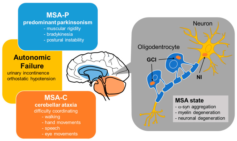
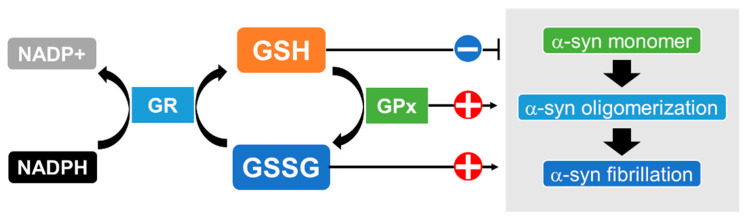
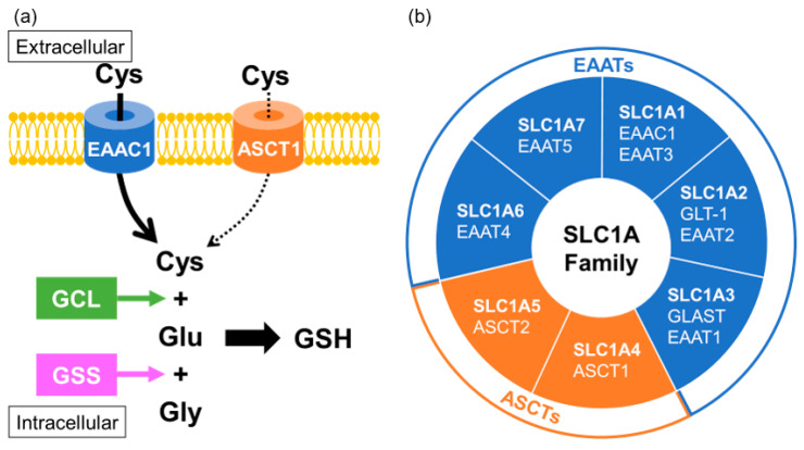
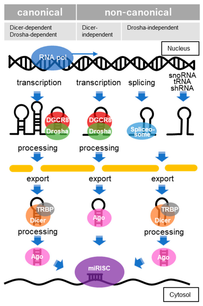
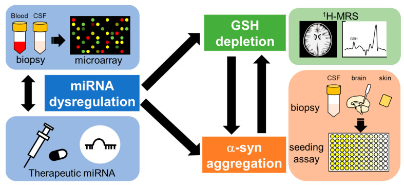
Multiple system atrophy (MSA) is a rare neurodegenerative disease characterized by parkinsonism, cerebellar impairment, and autonomic failure. Although the causes of MSA onset and progression remain uncertain, its pathogenesis may involve oxidative stress via the generation of excess reactive oxygen species and/or destruction of the antioxidant system. One of the most powerful antioxidants is glutathione, which plays essential roles as an antioxidant enzyme cofactor, cysteine-storage molecule, major redox buffer, and neuromodulator, in addition to being a key antioxidant in the central nervous system. Glutathione levels are known to be reduced in neurodegenerative diseases. In addition, genes regulating redox states have been shown to be post-transcriptionally modified by microRNA (miRNA), one of the most important types of non-coding RNA. miRNAs have been reported to be dysregulated in several diseases, including MSA. In this review, we focused on the relation between glutathione deficiency, miRNA dysregulation and oxidative stress and their close relation with MSA pathology.
Keywords: glutathione; microRNA; multiple system atrophy; neurodegenerative disease; oxidative stress; α-synuclein.
Conflict of interest statement
The authors declare no conflict of interest.
Figures
Similar articles
-
Multiple system atrophy of the cerebellar type: clinical state of the art.Mov Disord. 2014 Mar;29(3):294-304. doi: 10.1002/mds.25847. Epub 2014 Feb 24. Mov Disord. 2014. PMID: 24615754 Review.
-
Role of transcriptional control in multiple system atrophy.Neurobiol Aging. 2015 Jan;36(1):394-400. doi: 10.1016/j.neurobiolaging.2014.08.015. Epub 2014 Aug 19. Neurobiol Aging. 2015. PMID: 25218777 Review.
-
Is Multiple System Atrophy a Prion-like Disorder?Int J Mol Sci. 2021 Sep 18;22(18):10093. doi: 10.3390/ijms221810093. Int J Mol Sci. 2021. PMID: 34576255 Free PMC article. Review.
-
Human alpha-synuclein overexpressing MBP29 mice mimic functional and structural hallmarks of the cerebellar subtype of multiple system atrophy.Acta Neuropathol Commun. 2021 Apr 14;9(1):68. doi: 10.1186/s40478-021-01166-x. Acta Neuropathol Commun. 2021. PMID: 33853667 Free PMC article.
-
MicroRNAs Dysregulation and Metabolism in Multiple System Atrophy.Front Neurosci. 2019 Oct 17;13:1103. doi: 10.3389/fnins.2019.01103. eCollection 2019. Front Neurosci. 2019. PMID: 31680837 Free PMC article. Review.
Cited by
-
Bilirubin and Redox Stress in Age-Related Brain Diseases.Antioxidants (Basel). 2023 Jul 29;12(8):1525. doi: 10.3390/antiox12081525. Antioxidants (Basel). 2023. PMID: 37627520 Free PMC article. Review.
-
Decoding Parkinson's Disease: The interplay of cell death pathways, oxidative stress, and therapeutic innovations.Redox Biol. 2025 Jul 23;85:103787. doi: 10.1016/j.redox.2025.103787. Online ahead of print. Redox Biol. 2025. PMID: 40712453 Free PMC article. Review.
-
Natural Compounds and Glutathione: Beyond Mere Antioxidants.Antioxidants (Basel). 2023 Jul 18;12(7):1445. doi: 10.3390/antiox12071445. Antioxidants (Basel). 2023. PMID: 37507985 Free PMC article. Review.
-
Pharmacological inhibition of FABP7 by MF 6 counteracts cerebellum dysfunction in an experimental multiple system atrophy mouse model.Acta Pharmacol Sin. 2024 Jan;45(1):66-75. doi: 10.1038/s41401-023-01138-y. Epub 2023 Aug 21. Acta Pharmacol Sin. 2024. PMID: 37605049 Free PMC article.
References
Publication types
MeSH terms
Substances
Grants and funding
LinkOut - more resources
Full Text Sources
