Review
. 2022 Dec 1;23(23):15101.
doi: 10.3390/ijms232315101.

Trigeminal Sensory Supply Is Essential for Motor Recovery after Facial Nerve Injury

Affiliations
Review

Trigeminal Sensory Supply Is Essential for Motor Recovery after Facial Nerve Injury

Svenja Rink-Notzon et al. Int J Mol Sci. .

Abstract

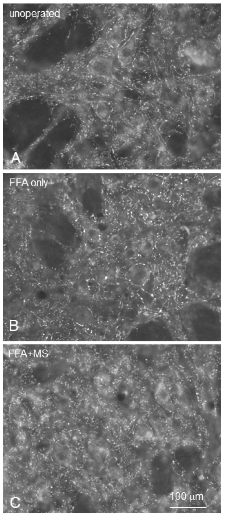
Recovery of mimic function after facial nerve transection is poor. The successful regrowth of regenerating motor nerve fibers to reinnervate their targets is compromised by (i) poor axonal navigation and excessive collateral branching, (ii) abnormal exchange of nerve impulses between adjacent regrowing axons, namely axonal crosstalk, and (iii) insufficient synaptic input to the axotomized facial motoneurons. As a result, axotomized motoneurons become hyperexcitable but unable to discharge. We review our findings, which have addressed the poor return of mimic function after facial nerve injuries, by testing the hypothesized detrimental component, and we propose that intensifying the trigeminal sensory input to axotomized and electrophysiologically silent facial motoneurons improves the specificity of the reinnervation of appropriate targets. We compared behavioral, functional, and morphological parameters after single reconstructive surgery of the facial nerve (or its buccal branch) with those obtained after identical facial nerve surgery, but combined with direct or indirect stimulation of the ipsilateral infraorbital nerve. We found that both methods of trigeminal sensory stimulation, i.e., stimulation of the vibrissal hairs and manual stimulation of the whisker pad, were beneficial for the outcome through improvement of the quality of target reinnervation and recovery of vibrissal motor performance.

Keywords: axotomy; facial nerve; morphological correlation; motion analysis; motoneuron; rat; recovery of function; stimulation; trigeminal nerve; vibrissal whisking.


Conflict of interest statement

The authors declare no conflict of interest.

Figures

Figure 7
Figure 7
(A): Schematic drawing illustrating the transection and suture site of the buccal branch of the facial nerve (Bn-n) and the resection of Ramus marginalis mandibulae of the rat facial nerve. (B): The large arrow indicates the site of transection of the buccal branch. Arrowheads point at the sites of tracer application in the superior and inferior buccolabial nerves. (CH): The rat brainstem 28 days after Bn-n. The photographs were produced by double exposure. The dorsomedial portion of the facial nucleus is indicated by an arrow. (C): The contralateral unlesioned lateral facial subnucleus with myotopic organization of the motoneurons, whose axons project into the superior buccolabial nerve [retrogradely labelled in white by Fluoro-Gold (FG)] and into the inferior buccolabial nerve (labelled in red by DiI). Most FG-labelled motoneurons are localized in the ventrolateral portion, and those labelled with DiI are in the dorsomedial part of the subnucleus. (D): The lesioned lateral facial subnucleus after Bn-n and application of FG to the superior, and DiI to the inferior buccolabial nerves. Note the complete lack of myotopic organization with the FG-labelled (white), DiI-labelled (red), and DiI + FG-labelled (arrowheads) motoneurons scattered throughout the entire lateral facial subnucleus. (E): The rat brainstem 28 days after Bn-n and excision of the ipsilateral infraorbital nerve with the contralateral unlesioned lateral facial subnucleus having myotopic organization. (F): The lesioned facial subnucleus 28 days after Bn-n and excision of the ipsilateral infraorbital nerve. (G): The rat brainstem 28 days after Bn-n and excision of the contralateral infraorbital nerve. The contralateral unlesioned lateral facial subnucleus has the normal myotopic distribution of the motoneurons. (H): The lateral facial subnucleus 28 days after Bn-n and excision of the contralateral infraorbital nerve; 50 μm vibratome sections. From Angelov et al. (1999) [90].
Figure 1
Figure 1
Video-based motion analysis of vibrissae motor performance on the left unoperated and right operated sides (OP-side) of the rat. Angles, angular velocity, and angular acceleration were measured during vibrissal protraction (a) and retraction (b). Note the significant change in angle from a sagittal line [Fr-Occ, which connects the frontally located tip of the nose (Fr) with the occipital bone (Occ)] during protraction and retraction on the intact side. The vibrissae on the operated side remain spastic (From Guntinas-Lichius et al., 2002) [59].
Figure 2
Figure 2
Schematic drawings illustrating the infratemporal portion of the facial nerve (Fn), and (A) the site of its transection and suture in one experimental rat group, as indicated by an arrow and (B) the close relationship between the peripheral fascicles of the Fn and those of the infraorbital nerve (IOn). The arrow shows the site of excision of the IOn in the experiments described in Section 4, demonstrating that afferent connections are important in improving functional recovery of whisking behavior and reducing polyinnervation of the motor endplates. The drawings are from Pavlov et al. (2008) [41].
Figure 3
Figure 3
(A) Schematic drawing of the extrinsic vibrissae muscles according to Dörfl (1982) [74]: α-δ: the four caudal hair follicles, the muscles slings of which “straddle” the five vibrissae rows (A1E1), T, transversus nasi muscle; L, levator labii superioris muscle; N, nasalis muscle; M, maxilolabialis muscle; O, orbit; S, septum intermusculare. Panels (B,C) show examples of a monoinnervated and a polyinnervated endplate, respectively, where superimposed stacks of confocal images of endplates in the levator labii superioris muscles of intact and surgically treated rats were visualized by staining the motor endplates with Alexa Fluor 488 α-bungarotoxin (green fluorescence), and immunostaining of the intramuscular axons for neuronal class III β-tubulin (Cy3 red fluorescence). Three axonal branches (arrows in (C)) reach the boundaries of the polyinnervated endplate delineated by the alpha-bungarotoxin staining. In contrast, the normally monoinnervated endplates in (B) are contacted by a single axon (empty arrows) with several preterminal rami, whilst in (C) the polyneuronally innervated endplate is contacted by three axonal branches. The drawing is from Sinis et al. (2009) [75].
Figure 4
Figure 4
Schematic drawings illustrating transection and end-to-end suture of the infratemporal portion of the right motor facial nerve (Fn-n; (A), lower arrow) and the sensory infraorbital branch of the trigeminal nerve, IOn-n ((A), upper arrow), adopted from (Dörfl 1985; Semba and Egger 1986) [66,76]. (B,C) Images of postoperative treatments: (B) The vibrissae on the left side of the face were trimmed to maximize vibrissal use on the operated, right side; (C) Manual stimulation of the whisker pad skin and musculature on the operated, right, side. (AC): From Bendella et al. (2011) [77].
Figure 5
Figure 5
(A): Schematic drawing indicating the injection site of the retrograde tracer Fast Blue (FB; syringe) into the whisker pad. (BE): Retrogradely labelled motoneuronal perikarya (blue) and synaptophysin-CY3 immunostaining of axosomatic nerve boutons in the intact facial nucleus (B), four months after Fn-n + Ion-n (C), after Fn-n + Ion-n + Vstim (D) and after Fn-n + Ion-n + Vstim + Mstim. FB-images were used to define “regions of interest” (RoIs) in each picture of the facial nucleus (ImageJ Software v1.38, NIH, Bethesda, MD, USA) through the following steps: (1) The dynamic range of the FB-images was maximized using gamma correction (γ = 0.2); (2) Images were sharpened by subtraction of a blurred copy (Gaussian blurring radius = 75 px); (3) Images were automatically thresholded using the Otsu algorithm to produce binary black and white images; (4) Motoneurons were included by selecting only FB-labelled areas with a value equal or greater than 500 µm2. (5) The resulting masks were used to measure the perimeter and area of the selected motoneurons. The perimeters were drawn and expanded in and out by 2 px, which generated RoIs from the closest perisomatic vicinity of the motoneurons with a width of 5 px (≈4 µm). All synaptophysin-positive profiles found within each of the predefined perisomatic RoIs of the thresholded images were counted, and the “numbers of perisomatic synapses per motoneuron” determined. From Bendella et al. (2011) [77].
Figure 6
Figure 6
Analysis of perisomatic puncta. Low-power confocal images (1-µm-thick optical slices) show the appearance of synaptophysin-positive puncta (arrows) around intact (A) and axotomized (B) facial motoneurons, identified by the retrograde labeling with FB. High-power confocal images used for counting: (C) intact and (D) axotomized motoneuronal cell bodies (*), four months after regeneration following facial and infraorbital nerve transection and end-to-end suture. Immunostained sections through the facial nucleus were examined under a fluorescence microscope. Stacks of images of 1 µm thickness were obtained on a TCS SP5 confocal microscope (Leica) using a 40 × oil immersion objective and digital resolution of 1024 × 1024 pixels. Four adjacent stacks (frame size, 115 × 115 µm) were obtained consecutively in a rostrocaudal direction to sample more motoneurons. One image per cell at the level of the largest cell body cross-sectional area was used to count the number of perisomatic puncta. Motoneurons were easily identified by the retrograde labeling with fast blue (FB). Areas and perimeters were measured using the Image Tool 2.0 software program (University of Texas, San Antonio, TX, USA). Linear density was calculated as the number of perisomatic puncta per unit length. Between 105 and 120 cells were analyzed per group and parameter. From Bendella et al. (2011) [77].
Figure 8
Figure 8
Rat brainstem 28 days after surgery on the buccal branch of the facial nerve (Bn). The lateral facial subnucleus, indicated by the pre-operative FG labelling, is in the left part of each picture. All photographs in the right column were produced by double exposure. (AC) Intact facial nucleus with preserved myotopic organization of the motoneurons. Employing the selective filters, we depicted all pre-operatively FG-labelled (A) and all postoperatively FB-labelled (B) motoneurons. (C) In the intact facial nucleus, the proportion of double-labelled (FG + FB, pink to bright purple in colour) motoneurons is about 90%. (DF) Lesioned facial nucleus 28 days after Bn-n. Whereas all preoperatively FG-labelled motoneurons are localized in the lateral facial subnucleus (D), those labelled postoperatively with FB are observed also in the intermediate facial subnucleus (E). Our quantitative estimates show that only about 27% of these FB-labelled motoneurons are double labelled (F) and belong to the original motoneuronal pool of the whisker pad. (GI). Lesioned facial nucleus of a rat 28 days after Bn-n + IOn-ipsi-ex. All preoperatively FG-labelled motoneurons are in the lateral facial subnucleus (G). The postoperatively FB-labelled motoneurons are found in the lateral and intermediate facial subnuclei (H). The double-exposure picture (I) is similar to that in F, showing that about 32% of the FB-labelled cells were also FG labelled. (JL). Lesioned facial nucleus of rat 28 days after Bn-n +IOn-contra-ex. All preoperatively FG-labelled motoneurons are in the lateral facial subnucleus (J) and some postoperatively FB-labelled cells are found in the intermediate facial subnucleus (K). Our counts show that after this type of combined surgery, the proportion of the double-labelled motoneurons (L) increased significantly to 41%. 50 µm thick vibratome sections; the scale bar indicates 100 μm. From Skouras et al. (2002) [91].
Figure 9
Figure 9
Immunostaining for synaptophysin in 30-μm thick vibratome sections from the facial nucleus in intact rats (A), in rats 2 months after Fn-n and handling (Fn-n + handling; (B)) and in rats that received Mstim of the vibrissal muscles after Fn-n (Fn-n + Mstim; (C)). Note the clearly discernible numerous puncta within the neuropil and around motoneuronal cell bodies in the facial nucleus representing synaptic terminals. From Pavlov et al. (2008) [41].

Similar articles

Cited by

References

    1. Markiewicz M.R., Callahan N., Miloro M. Management of Traumatic Trigeminal and Facial Nerve Injuries. Oral Maxillofac. Surg. Clin. N. Am. 2021;33:381–405. doi: 10.1016/j.coms.2021.04.009. - DOI - PubMed
    1. Holland N.J., Bernstein J.M. Bell’s palsy. BMJ Clin. Evid. 2014;2014:1204. - PMC - PubMed
    1. Zhang W., Xu L., Luo T., Wu F., Zhao B., Li X. The etiology of Bell’s palsy: A review. J. Neurol. 2020;267:1896–1905. doi: 10.1007/s00415-019-09282-4. - DOI - PMC - PubMed
    1. Anonsen C.K., Trachy R.E., Hibbert J., Cummings C.W. Assessment of facial reinnervation by use of chronic electromyographic monitoring. Otolaryngol. Neck Surg. Off. J. Am. Acad. Otolaryngol. Neck Surg. 1986;94:32–36. doi: 10.1177/019459988609400105. - DOI - PubMed
    1. Ferreira M.C., Besteiro J.M., Tuma Júnior P. Results of reconstruction of the facial nerve. Microsurgery. 1994;15:5–8. doi: 10.1002/micr.1920150104. - DOI - PubMed