Impact of Sulfated Hyaluronan on Bone Metabolism in Diabetic Charcot Neuroarthropathy and Degenerative Arthritis
- PMID: 36499493
- PMCID: PMC9737841
- DOI: 10.3390/ijms232315146
Impact of Sulfated Hyaluronan on Bone Metabolism in Diabetic Charcot Neuroarthropathy and Degenerative Arthritis
Abstract
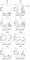
Bone in diabetes mellitus is characterized by an altered microarchitecture caused by abnormal metabolism of bone cells. Together with diabetic neuropathy, this is associated with serious complications including impaired bone healing culminating in complicated fractures and dislocations, especially in the lower extremities, so-called Charcot neuroarthropathy (CN). The underlying mechanisms are not yet fully understood, and treatment of CN is challenging. Several in vitro and in vivo investigations have suggested positive effects on bone regeneration by modifying biomaterials with sulfated glycosaminoglycans (sGAG). Recent findings described a beneficial effect of sGAG for bone healing in diabetic animal models compared to healthy animals. We therefore aimed at studying the effects of low- and high-sulfated hyaluronan derivatives on osteoclast markers as well as gene expression patterns of osteoclasts and osteoblasts from patients with diabetic CN compared to non-diabetic patients with arthritis at the foot and ankle. Exposure to sulfated hyaluronan (sHA) derivatives reduced the exaggerated calcium phosphate resorption as well as the expression of genes associated with bone resorption in both groups, but more pronounced in patients with CN. Moreover, sHA derivatives reduced the release of pro-inflammatory cytokines in osteoclasts of patients with CN. The effects of sHA on osteoblasts differed only marginally between patients with CN and non-diabetic patients with arthritis. These results suggest balancing effects of sHA on osteoclastic bone resorption parameters in diabetes.
Keywords: Charcot neuroarthropathy; ankle arthritis; diabetes mellitus; osteoblasts; osteoclasts; sulfated hyaluronan.
Conflict of interest statement
The authors declare no conflict of interest.
Figures


Similar articles
-
Measurement of markers of osteoclast and osteoblast activity in patients with acute and chronic diabetic Charcot neuroarthropathy.Diabet Med. 1997 Jul;14(7):527-31. doi: 10.1002/(SICI)1096-9136(199707)14:7<527::AID-DIA404>3.0.CO;2-Q. Diabet Med. 1997. PMID: 9223389
-
Sulfated hyaluronan improves bone regeneration of diabetic rats by binding sclerostin and enhancing osteoblast function.Biomaterials. 2016 Jul;96:11-23. doi: 10.1016/j.biomaterials.2016.04.013. Epub 2016 Apr 21. Biomaterials. 2016. PMID: 27131598
-
Diabetic charcot neuroarthropathy of the foot and ankle with osteomyelitis.Clin Podiatr Med Surg. 2014 Oct;31(4):487-92. doi: 10.1016/j.cpm.2013.12.001. Clin Podiatr Med Surg. 2014. PMID: 25281510 Review.
-
Autoantibodies to post-translationally modified type I and II collagen in Charcot neuroarthropathy in subjects with type 2 diabetes mellitus.Diabetes Metab Res Rev. 2017 Feb;33(2). doi: 10.1002/dmrr.2839. Epub 2016 Oct 5. Diabetes Metab Res Rev. 2017. PMID: 27454862
-
Diabetes Mellitus: An Overview in Relationship to Charcot Neuroarthropathy.Clin Podiatr Med Surg. 2022 Oct;39(4):535-542. doi: 10.1016/j.cpm.2022.05.001. Epub 2022 Aug 6. Clin Podiatr Med Surg. 2022. PMID: 36180186 Review.
Cited by
-
Cellular response of advanced triple cultures of human osteocytes, osteoblasts and osteoclasts to high sulfated hyaluronan (sHA3).Mater Today Bio. 2024 Feb 22;25:101006. doi: 10.1016/j.mtbio.2024.101006. eCollection 2024 Apr. Mater Today Bio. 2024. PMID: 38445011 Free PMC article.
-
[Progress in clinical diagnosis and treatment of diabetic Charcot neuroarthropathy of foot and ankle].Zhongguo Xiu Fu Chong Jian Wai Ke Za Zhi. 2023 Nov 15;37(11):1438-1443. doi: 10.7507/1002-1892.202307068. Zhongguo Xiu Fu Chong Jian Wai Ke Za Zhi. 2023. PMID: 37987057 Free PMC article. Chinese.
-
Therapeutics of Charcot neuroarthropathy and pharmacological mechanisms: A bone metabolism perspective.Front Pharmacol. 2023 Apr 12;14:1160278. doi: 10.3389/fphar.2023.1160278. eCollection 2023. Front Pharmacol. 2023. PMID: 37124200 Free PMC article. Review.
References
-
- Ahluwalia R., Bilal A., Petrova N., Boddhu K., Manu C., Vas P., Bates M., Corcoran B., Reichert I., Mulholland N., et al. The Role of Bone Scintigraphy with SPECT/CT in the Characterization and Early Diagnosis of Stage 0 Charcot Neuroarthropathy. J. Clin. Med. 2020;9:4123. doi: 10.3390/jcm9124123. - DOI - PMC - PubMed
-
- Rammelt S. Management of Acute Hindfoot Fractures in Diabetics. Apr 16, 2016. [(accessed on 13 July 2022)]. Available online: https://link.springer.com/chapter/10.1007/978-3-319-27623-6_7. - DOI
-
- Amadou C., Carlier A., Amouyal C., Bourron O., Aubert C., Couture T., Fourniols E., Ha Van G., Rouanet S., Hartemann A. Five-year mortality in patients with diabetic foot ulcer during 2009–2010 was lower than expected. Diabetes Metab. 2020;46:230–235. doi: 10.1016/j.diabet.2019.04.010. - DOI - PubMed
MeSH terms
Substances
Grants and funding
LinkOut - more resources
Full Text Sources
Medical

