

CD200 as a Potential New Player in Inflammation during Rotator Cuff Tendon Injury/Repair: An In Vitro Model
- PMID: 36499497
- PMCID: PMC9738060
- DOI: 10.3390/ijms232315165
CD200 as a Potential New Player in Inflammation during Rotator Cuff Tendon Injury/Repair: An In Vitro Model
Abstract
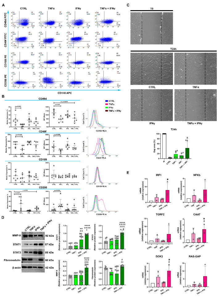
Rotator cuff tendon (RCT) disease results from multifactorial mechanisms, in which inflammation plays a key role. Pro-inflammatory cytokines and tendon stem cell/progenitor cells (TSPCs) have been shown to participate in the inflammatory response. However, the underlying molecular mechanism is still not clear. In this study, flow cytometry analyses of different subpopulations of RCT-derived TSPCs demonstrate that after three days of administration, TNFα alone or in combination with IFNγ significantly decreases the percentage of CD146+CD49d+ and CD146+CD49f+ but not CD146+CD109+ TSPCs populations. In parallel, the same pro-inflammatory cytokines upregulate the expression of CD200 in the CD146+ TSPCs population. Additionally, the TNFα/IFNγ combination modulates the protein expression of STAT1, STAT3, and MMP9, but not fibromodulin. At the gene level, IRF1, CAAT (CAAT/EBPbeta), and DOK2 but not NF-κb, TGRF2 (TGFBR2), and RAS-GAP are modulated. In conclusion, although our study has several important limitations, the results highlight a new potential role of CD200 in regulating inflammation during tendon injuries. In addition, the genes analyzed here might be new potential players in the inflammatory response of TSPCs.
Keywords: C/EBPbeta; CD146; CD200; DOK2; IFNγ; IRF1; TNFα; inflammation; rotator cuff disease; tendon stem cells; tendons.
Conflict of interest statement
The authors declare no conflict of interest.
Figures

Similar articles
-
Various Strategies of Tendon Stem/Progenitor Cell Reprogramming for Tendon Regeneration.Int J Mol Sci. 2024 Nov 1;25(21):11745. doi: 10.3390/ijms252111745. Int J Mol Sci. 2024. PMID: 39519296 Free PMC article. Review.
-
Decellularized Human Umbilical Cord Wharton Jelly Scaffold Improves Tendon Regeneration in a Rabbit Rotator Cuff Tendon Defect Model.Am J Sports Med. 2022 Feb;50(2):371-383. doi: 10.1177/03635465211055722. Epub 2021 Nov 5. Am J Sports Med. 2022. PMID: 34739346
-
Allogenous tendon stem/progenitor cells in silk scaffold for functional shoulder repair.Cell Transplant. 2012;21(5):943-58. doi: 10.3727/096368911X627453. Epub 2012 Mar 8. Cell Transplant. 2012. PMID: 22405331
-
The interaction between human rotator cuff tendon and subacromial bursal tissue in co-culture.J Shoulder Elbow Surg. 2021 Jul;30(7):1494-1502. doi: 10.1016/j.jse.2020.09.025. Epub 2020 Nov 13. J Shoulder Elbow Surg. 2021. PMID: 33197595
-
[Progress on tendon-to-bone healing after rotator cuff repair].Zhongguo Gu Shang. 2018 Dec 25;31(12):1172-1179. doi: 10.3969/j.issn.1003-0034.2018.12.020. Zhongguo Gu Shang. 2018. PMID: 30583662 Review. Chinese.
Cited by
-
Global research trends and hotspots on tendon-derived stem cell: a bibliometric visualization study.Front Bioeng Biotechnol. 2024 Jan 8;11:1327027. doi: 10.3389/fbioe.2023.1327027. eCollection 2023. Front Bioeng Biotechnol. 2024. PMID: 38260747 Free PMC article.
-
Delivery of GAS5 by LNP promotes tendon-bone healing in rotator cuff injury.Bioact Mater. 2025 Jun 24;51:758-773. doi: 10.1016/j.bioactmat.2025.06.009. eCollection 2025 Sep. Bioact Mater. 2025. PMID: 40656649 Free PMC article.
-
The combination of hyaluronic acids and collagen boosts human Achilles tendon-derived cell escape from inflammation and matrix remodeling in vitro.Inflamm Res. 2025 Jan 7;74(1):4. doi: 10.1007/s00011-024-01975-5. Inflamm Res. 2025. PMID: 39762639
-
In Vitro CO-Releasing and Antioxidant Properties of Sulfonamide-Based CAI-CORMs in a H2O2-Stimulated Human Achilles Tendon-Derived Cell Model.Molecules. 2025 Jan 28;30(3):593. doi: 10.3390/molecules30030593. Molecules. 2025. PMID: 39942697 Free PMC article.
References
MeSH terms
Substances
LinkOut - more resources
Full Text Sources
Medical
Research Materials
Miscellaneous
