

Associations between Maternal Risk Factors and Intrinsic Placental and Fetal Brain Functional Properties in Congenital Heart Disease
- PMID: 36499505
- PMCID: PMC9738149
- DOI: 10.3390/ijms232315178
Associations between Maternal Risk Factors and Intrinsic Placental and Fetal Brain Functional Properties in Congenital Heart Disease
Abstract
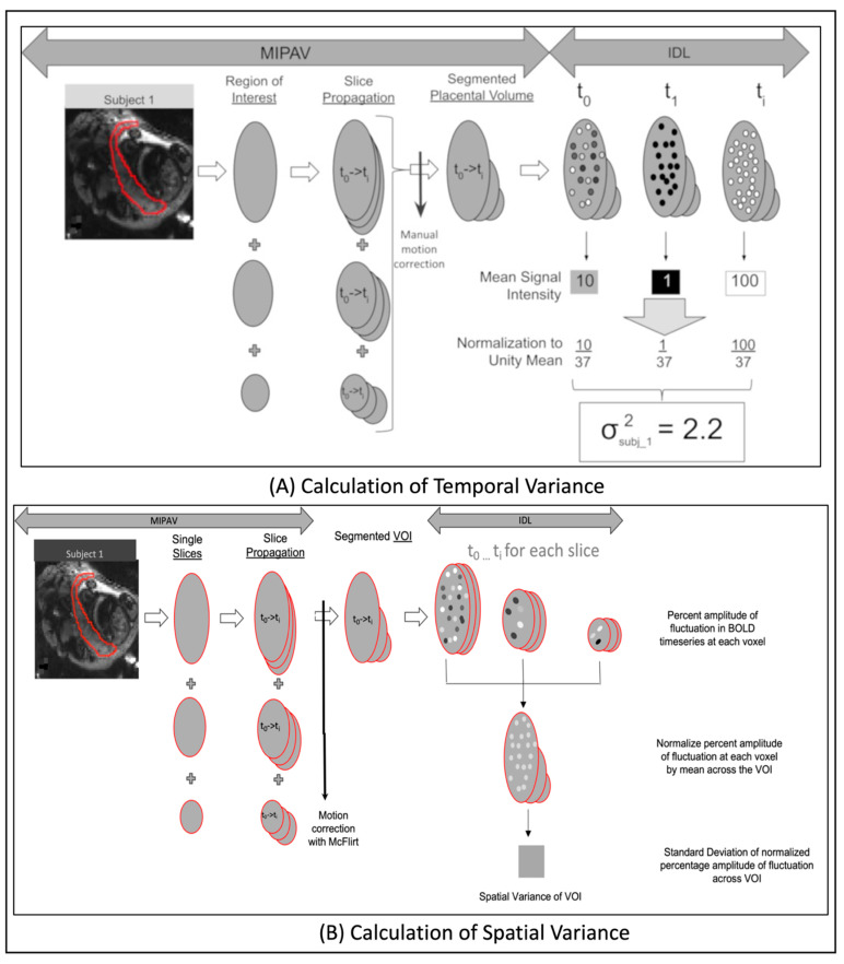
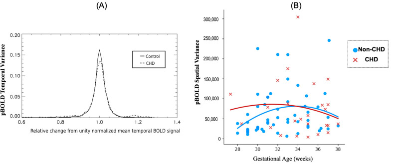
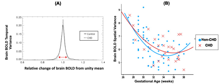
The relationship between maternal risk factors (MRFs) (particularly pre-gravid obesity, diabetes, and hypertension) and congenital heart disease (CHD) to placental and fetal brain outcomes is poorly understood. Here, we tested the hypothesis that MRF and CHD would be associated with reduced intrinsic placental and fetal brain function using a novel non-invasive technique. Pregnant participants with and without MRF and fetal CHD were prospectively recruited and underwent feto-placental MRI. Using intrinsic properties of blood oxygen level dependent imaging (BOLD) we quantified spatiotemporal variance of placenta and fetal brain. MRFs and CHD were correlated with functional characteristics of the placenta and fetal brain. Co-morbid MRF (hypertension, diabetes, and obesity) reduced spatiotemporal functional variance of placenta and fetal brain (p < 0.05). CHD predicted reduced fetal brain temporal variance compared to non-CHD (p < 0.05). The presence of both MRF and CHD was associated with reduced intrinsic pBOLD temporal variance (p = 0.047). There were no significant interactions of MRFs and CHD status on either temporal or spatial variance of intrinsic brain BOLD. MRF and CHD reduced functional characteristic of placenta and brain in fetuses. MRF modification and management during pregnancy may have the potential to not only provide additional risk stratification but may also improve neurodevelopmental outcomes.
Keywords: fetal brain resting BOLD signal; maternal diabetes; maternal hypertension; placenta.
Conflict of interest statement
The authors declare no conflict of interest.
Figures
Similar articles
-
Interdependence of placenta and fetal cardiac development.Prenat Diagn. 2024 Jun;44(6-7):846-855. doi: 10.1002/pd.6572. Epub 2024 Apr 27. Prenat Diagn. 2024. PMID: 38676696 Free PMC article. Review.
-
Hemodynamic Responses of the Placenta and Brain to Maternal Hyperoxia in Fetuses with Congenital Heart Disease by Using Blood Oxygen-Level Dependent MRI.Radiology. 2020 Jan;294(1):141-148. doi: 10.1148/radiol.2019190751. Epub 2019 Nov 5. Radiology. 2020. PMID: 31687920 Free PMC article.
-
T2*-Relaxometry MRI to Assess Third Trimester Placental and Fetal Brain Oxygenation and Placental Characteristics in Healthy Fetuses and Fetuses With Congenital Heart Disease.J Magn Reson Imaging. 2025 Mar;61(3):1246-1255. doi: 10.1002/jmri.29498. Epub 2024 Jul 12. J Magn Reson Imaging. 2025. PMID: 38994701 Free PMC article.
-
Neuroplacentology in congenital heart disease: placental connections to neurodevelopmental outcomes.Pediatr Res. 2022 Mar;91(4):787-794. doi: 10.1038/s41390-021-01521-7. Epub 2021 Apr 16. Pediatr Res. 2022. PMID: 33864014 Free PMC article. Review.
-
3-D volumetric MRI evaluation of the placenta in fetuses with complex congenital heart disease.Placenta. 2015 Sep;36(9):1024-30. doi: 10.1016/j.placenta.2015.06.013. Epub 2015 Jul 6. Placenta. 2015. PMID: 26190037 Free PMC article.
Cited by
-
Interdependence of placenta and fetal cardiac development.Prenat Diagn. 2024 Jun;44(6-7):846-855. doi: 10.1002/pd.6572. Epub 2024 Apr 27. Prenat Diagn. 2024. PMID: 38676696 Free PMC article. Review.
-
Altered In Utero Metabolic Brain Trajectories in CHD: Going Beyond Fetal Brain Structure and Physiology.J Am Coll Cardiol. 2023 Oct 17;82(16):1624-1627. doi: 10.1016/j.jacc.2023.08.039. J Am Coll Cardiol. 2023. PMID: 37821173 Free PMC article. No abstract available.
-
Postnatal Cerebral Hemodynamics and Placental Vascular Malperfusion Lesions in Neonates With Congenital Heart Disease.Pediatr Neurol. 2024 Jul;156:72-78. doi: 10.1016/j.pediatrneurol.2024.03.028. Epub 2024 Apr 6. Pediatr Neurol. 2024. PMID: 38733857 Free PMC article.
References
-
- Veerbeek J., Nikkels P., Torrance H., Gravesteijn J., Uiterweer E.P., Derks J., Koenen S., Visser G., Van Rijn B., Franx A. Placental pathology in early intrauterine growth restriction associated with maternal hypertension. Placenta. 2014;35:696–701. doi: 10.1016/j.placenta.2014.06.375. - DOI - PubMed
MeSH terms
Grants and funding
LinkOut - more resources
Full Text Sources
Medical
Miscellaneous
