Review
. 2022 Dec 2;23(23):15186.
doi: 10.3390/ijms232315186.

Endoplasmic Reticulum Stress Signaling and Neuronal Cell Death

Affiliations
Review

Endoplasmic Reticulum Stress Signaling and Neuronal Cell Death

Adalberto Merighi et al. Int J Mol Sci. .

Abstract

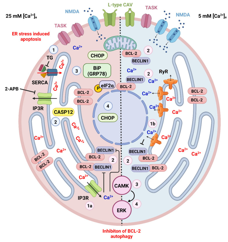
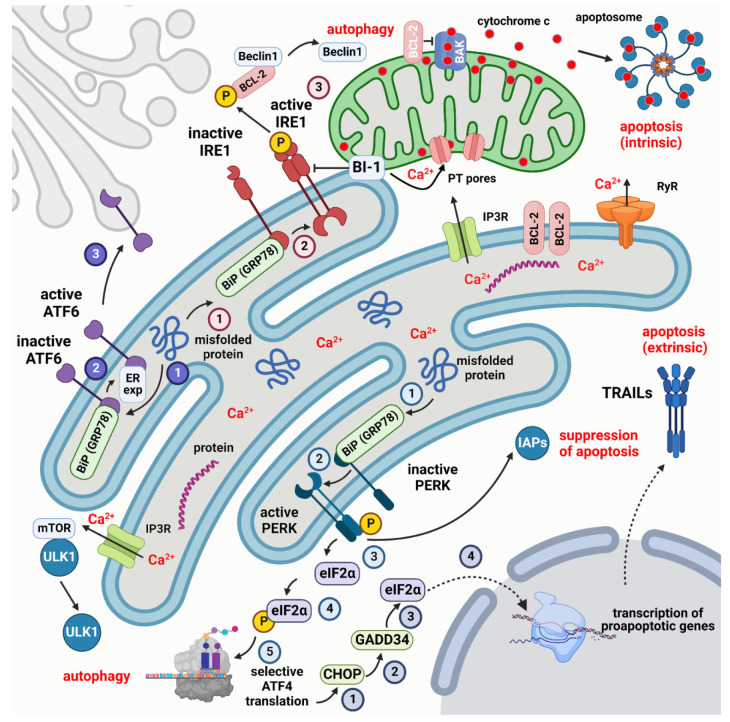
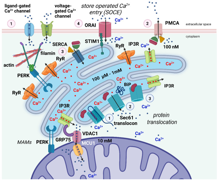
Besides protein processing, the endoplasmic reticulum (ER) has several other functions such as lipid synthesis, the transfer of molecules to other cellular compartments, and the regulation of Ca2+ homeostasis. Before leaving the organelle, proteins must be folded and post-translationally modified. Protein folding and revision require molecular chaperones and a favorable ER environment. When in stressful situations, ER luminal conditions or chaperone capacity are altered, and the cell activates signaling cascades to restore a favorable folding environment triggering the so-called unfolded protein response (UPR) that can lead to autophagy to preserve cell integrity. However, when the UPR is disrupted or insufficient, cell death occurs. This review examines the links between UPR signaling, cell-protective responses, and death following ER stress with a particular focus on those mechanisms that operate in neurons.

Keywords: apoptosis; autophagy; cerebellar granule cells; endoplasmic reticulum; endoplasmic reticulum stress; unfolded protein response.


Conflict of interest statement

The authors declare no conflict of interest. The funders had no role in the design of the study; in the collection, analyses, or interpretation of data; in the writing of the manuscript; or in the decision to publish the results.

Figures

Figure 3
Figure 3
ER stress and cell death in the CGCs. Apoptotic and autophagic mechanisms are dependent on the cytosolic and ER intraluminal concentration of Ca2+. These, in turn, are related to the culture conditions in high (on the left represented with pink cytoplasm) or physiological (on the right represented with azure cytoplasm) extracellular K+. Different cultural conditions lead to distinct patterns of expression of plasmalemmal receptors, particularly TASK channels that contribute to the resting membrane potential, L-type Ca2+ channels that were demonstrated to interact with RyRs in CGCs [232], and NMDA glutamate receptors. Several drugs have been used to induce ER stress in CGCs (see the main text) cultured in 25 mM [K+]e eventually leading to apoptosis. The figure shows the cellular events following thapsigargin (TG—numbered azure circles), a blocker of SERCA. With SERCA block, the influx of Ca2+ into the ER lumen stops, leading to the activation of CASP12 and the expression of the UPR molecules BiP(GRP78), phosphorylated eIF2α and CHOP, the latter eventually being translocated to the nucleus and promoting the transcription of proapoptotic genes. ER Ca2+ controls cellular homeostasis through two different mechanisms mediated by IP3Rs (1a—pink circle) or RyRs (1b—pink circle). The outflow of Ca2+ from the ER leads to an increase in the cytoplasmic concentration of the ion with the phosphorylation of BCL-2 and its dissociation from beclin1, thereby inhibiting BCL-2 autophagy through the activation of the CAMK-ERK pathway (pink circles 2–4) [128]. This mechanism operates in both high and physiological [K+]e. Created with Biorender.com (www.biorender.com, accessed on 27 November 2022).
Figure 1
Figure 1
Simplified scheme of the UPR response and its relation to autophagy and apoptosis. The three main sensors that regulate the UPR are PERK, IRE1, and ATF6. Their respective pathways are indicated by numbered circles in light blue, dark blue, and pink. PERK, IRE1, and ATF6 are maintained in an inactive state by binding to the chaperone BiP (GRP78). Misfolded proteins promote the dissociation of BiP (GRP78) and the activation of the three sensors. Active PERK, in turn, phosphorylates eIF2α to start the selective translation of autophagic proteins by ATF4. ATF4 can also activate CHOP that acts on GADD34 to dephosphorylate eIF2α promoting the transcription of proapoptotic genes among which TRAILs that activate extrinsic apoptosis. Active PERK can, however, inhibit apoptosis by binding to IAPs. Once freed from BiP (GRP78), IRE1 dephosphorylates cytoplasmic BCL-2 to release Beclin1 and trigger autophagy. The dissociation of ATF6 from BiP (GRP78) leads to binding to an ER export motif that starts the translocation of ATF6 to the Golgi apparatus. The MAM protein BI-1 controls the Ca2+ uptake in mitochondria and intrinsic apoptosis, with the intervention of the pro- (BAK, BAX) and antiapoptotic proteins (BCL-2) of the BCL-2 family. Created with Biorender.com (www.biorender.com, accessed on 27 November 2022).
Figure 2
Figure 2
Simplified scheme of Ca2+ dynamics inside the cell and its relation to ER stress. In excitable cells such as neurons, Ca2+ ions enter the cell through ligand-gated and voltage-gated channels (1—pink circle). The Ca2+ gradient across the cell membrane is actively maintained by the PMCA pump (2—pink circle). The ER is the main Ca2+-storing organelle. Ca2+ is transported against a steep concentration gradient into the lumen of the ER via the SERCA pump (3—pink circle). Upon ligand stimulation, Ca2+ ions are released from the reticulum into the cytosol via the IP3Rs and RyRs to activate Ca2+-dependent proteins and mediate stimulus-response signaling. The refilling of intracellular Ca2+ stores by extracellular Ca2+ occurs via SOCE (4—pink circle) by close apposition of the transmembrane proteins STIM1 and ORAI. Filamin A, an actin-binding protein, interacts with PERK independently from the UPR. This interaction seems to be crucial for the formation of juxtapositions of the ER membrane with the plasma membrane, the proximity of the two membranes being a precondition for SOCE to occur. Protein translocation across the ER membrane is mediated via the Sec61 translocon (1—light blue circle). At the end of the translocation process, the Sec61 channel mediates the efflux of Ca2+ from the ER to the cytosol resulting in a reduction in [Ca2+]ER (2—light blue circle). The ER chaperone BiP binds to the translocon to close it and block the Ca2+ efflux from the ER (3-light blue circle). Efficient Ca2+ flux from the ER to mitochondria is mediated via IP3Rs, the voltage-dependent anion channel 1 (VDAC1), which is located at the outer mitochondrial membrane, and mitochondrial Ca2+ uniporter 1 (MCU1), located at the inner mitochondrial membrane. The IP3Rs–VDAC1 interaction is facilitated by the chaperone GRP75, which is enriched in MAMs. Created with biorender.com (www.biorender.com, accessed on 27 November 2022).

Similar articles

Cited by

References

    1. Porter K.R., Claude A., Fullam E.F. A study of tissue culture cells by electron microscopy: Methods and preliminary observations. J. Exp. Med. 1945;81:233–246. doi: 10.1084/jem.81.3.233. - DOI - PMC - PubMed
    1. Palade G.E., Porter K.R. Studies on the endoplasmic reticulum. I. Its identification in cells in situ. J. Exp. Med. 1954;100:641–656. doi: 10.1084/jem.100.6.641. - DOI - PMC - PubMed
    1. Schwarz D.S., Blower M.D. The endoplasmic reticulum: Structure, function, and response to cellular signaling. Cell. Mol. Life Sci. 2016;73:79–94. doi: 10.1007/s00018-015-2052-6. - DOI - PMC - PubMed
    1. Nunes P., Demaurex N. Redox regulation of store-operated Ca2+ entry. Antioxid. Redox Signal. 2014;21:915–932. doi: 10.1089/ars.2013.5615. - DOI - PMC - PubMed
    1. Costa-Mattioli M., Sossin W.S., Klann E., Sonenberg N. Translational control of long-lasting synaptic plasticity and memory. Neuron. 2009;61:10–26. doi: 10.1016/j.neuron.2008.10.055. - DOI - PMC - PubMed

MeSH terms

Substances

Grants and funding

LinkOut - more resources