Tolerogenic Dendritic Cells Induce Apoptosis-Independent T Cell Hyporesponsiveness of SARS-CoV-2-Specific T Cells in an Antigen-Specific Manner
- PMID: 36499533
- PMCID: PMC9740551
- DOI: 10.3390/ijms232315201
Tolerogenic Dendritic Cells Induce Apoptosis-Independent T Cell Hyporesponsiveness of SARS-CoV-2-Specific T Cells in an Antigen-Specific Manner
Abstract
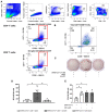
Although the global pandemic caused by the novel severe acute respiratory syndrome coronavirus 2 (SARS-CoV-2) is still ongoing, there are currently no specific and highly efficient drugs for COVID-19 available, particularly in severe cases. Recent findings demonstrate that severe COVID-19 disease that requires hospitalization is associated with the hyperactivation of CD4+ and CD8+ T cell subsets. In this study, we aimed to counteract this high inflammatory state by inducing T-cell hyporesponsiveness in a SARS-CoV-2-specific manner using tolerogenic dendritic cells (tolDC). In vitro-activated SARS-CoV-2-specific T cells were isolated and stimulated with SARS-CoV-2 peptide-loaded monocyte-derived tolDC or with SARS-CoV-2 peptide-loaded conventional (conv) DC. We demonstrate a significant decrease in the number of interferon (IFN)-γ spot-forming cells when SARS-CoV-2-specific T cells were stimulated with tolDC as compared to stimulation with convDC. Importantly, this IFN-γ downmodulation in SARS-CoV-2-specific T cells was antigen-specific, since T cells retain their capacity to respond to an unrelated antigen and are not mediated by T cell deletion. Altogether, we have demonstrated that SARS-CoV-2 peptide-pulsed tolDC induces SARS-CoV-2-specific T cell hyporesponsiveness in an antigen-specific manner as compared to stimulation with SARS-CoV-2-specific convDC. These observations underline the clinical potential of tolDC to correct the immunological imbalance in the critically ill.
Keywords: T cell apoptosis; antigen specificity; severe COVID-19; tolerance; tolerogenic dendritic cells.
Conflict of interest statement
The authors declare no conflict of interest.
Figures


Similar articles
-
Immunomodulatory Effects of 1,25-Dihydroxyvitamin D3 on Dendritic Cells Promote Induction of T Cell Hyporesponsiveness to Myelin-Derived Antigens.J Immunol Res. 2016;2016:5392623. doi: 10.1155/2016/5392623. Epub 2016 Sep 14. J Immunol Res. 2016. PMID: 27703987 Free PMC article.
-
Foxp3+ CD4+ regulatory T cells control dendritic cells in inducing antigen-specific immunity to emerging SARS-CoV-2 antigens.PLoS Pathog. 2021 Dec 9;17(12):e1010085. doi: 10.1371/journal.ppat.1010085. eCollection 2021 Dec. PLoS Pathog. 2021. PMID: 34882757 Free PMC article.
-
Targeting of tolerogenic dendritic cells to heat-shock proteins in inflammatory arthritis.J Transl Med. 2019 Nov 14;17(1):375. doi: 10.1186/s12967-019-2128-4. J Transl Med. 2019. PMID: 31727095 Free PMC article.
-
Perspective of HLA-G Induced Immunosuppression in SARS-CoV-2 Infection.Front Immunol. 2021 Dec 6;12:788769. doi: 10.3389/fimmu.2021.788769. eCollection 2021. Front Immunol. 2021. PMID: 34938296 Free PMC article. Review.
-
Cellular immune states in SARS-CoV-2-induced disease.Front Immunol. 2022 Nov 23;13:1016304. doi: 10.3389/fimmu.2022.1016304. eCollection 2022. Front Immunol. 2022. PMID: 36505442 Free PMC article. Review.
Cited by
-
Physiological Oxygen Levels in the Microenvironment Program Ex Vivo-Generated Conventional Dendritic Cells Toward a Tolerogenic Phenotype.Cells. 2025 May 18;14(10):736. doi: 10.3390/cells14100736. Cells. 2025. PMID: 40422239 Free PMC article.
References
-
- Ganesh B., Rajakumar T., Malathi M., Manikandan N., Nagaraj J., Santhakumar A., Elangovan A., Malik Y.S. Epidemiology and pathobiology of SARS-CoV-2 (COVID-19) in comparison with SARS, MERS: An updated overview of current knowledge and future perspectives. Clin. Epidemiol. Glob. Health. 2021;10:100694. doi: 10.1016/j.cegh.2020.100694. - DOI - PMC - PubMed
-
- World Health Organization . WHO Coronavirus (COVID-19) Dashboard. WHO; Geneva, Switzerland: 2021.
MeSH terms
Substances
Grants and funding
LinkOut - more resources
Full Text Sources
Medical
Research Materials
Miscellaneous

