. 2022 Dec 2;23(23):15219.
doi: 10.3390/ijms232315219.

Regulation of Prepro-NeuropeptideW/B and Its Receptor in the Heart of ZDF Rats: An Animal Model of Type II DM

Affiliations

Regulation of Prepro-NeuropeptideW/B and Its Receptor in the Heart of ZDF Rats: An Animal Model of Type II DM

Shashank Pandey et al. Int J Mol Sci. .

Abstract

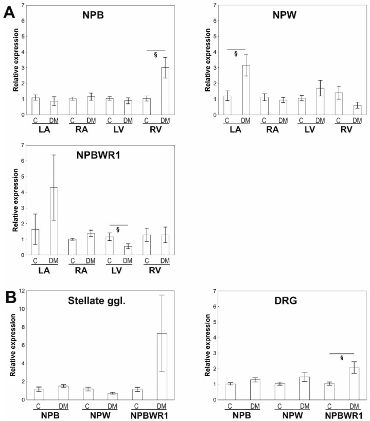
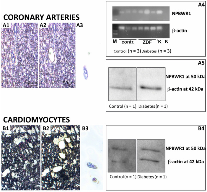
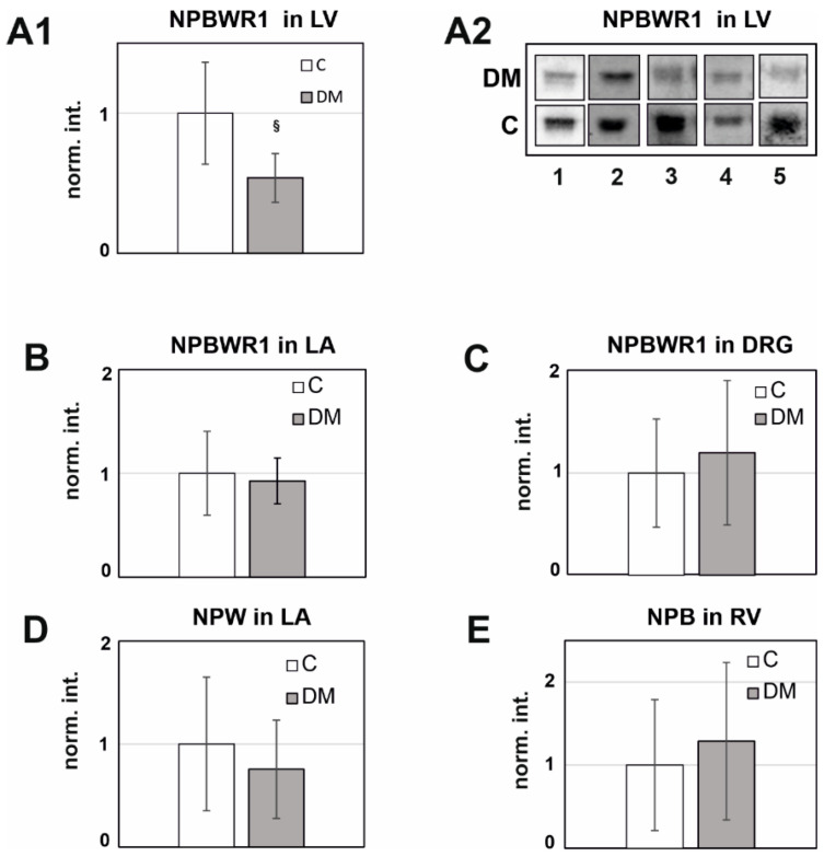
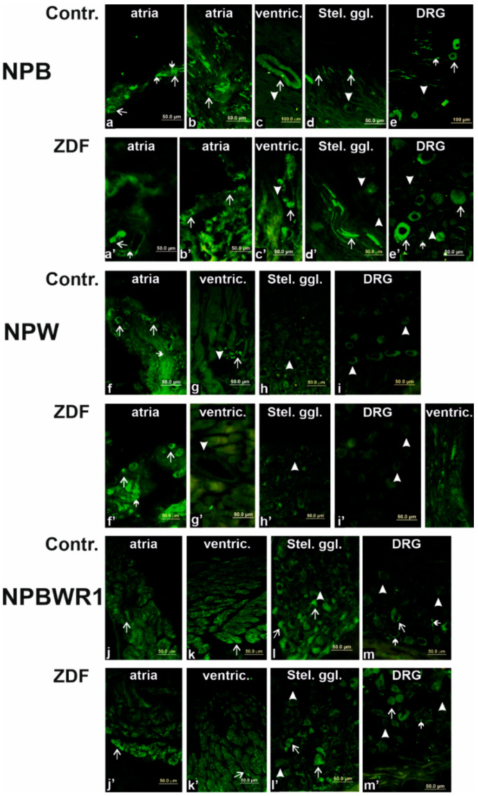
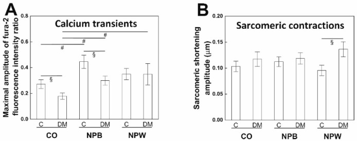
Neuropeptide B (NPB) and neuropeptide W (NPW) are neuropeptides, which constitute NPB/W signaling systems together with G-protein coupled receptors NPBWR1. The location and function of NPB/W signaling systems have been predominantly detected and mapped within the CNS, including their role in the modulation of inflammatory pain, neuroendocrine functions, and autonomic nervous systems. The aim of the study is to investigate the impact of diabetes on the neuropeptide B/W signaling system in different heart compartments and neurons which innervates it. In the RT-qPCR analysis, we observed the upregulation of mRNA for preproNPB in RV, for preproNPW in LA, and for NPBWR1 in DRG in diabetic rats. On the contrary, the expression of mRNA for NPBWR1 was downregulated in LV in diabetic rats. In the WB analysis, significant downregulation of NPBWR1 in LV (0.54-fold, p = 0.046) in diabetic rats was observed at the proteomic level. The presence of NPBWR1 was also confirmed in a dissected LCM section of cardiomyocytes and coronary arteries. The positive inotropic effect of NPW described on the diabetic cardiomyocytes in vitro could point to a possible therapeutic target for compensation of the contractile dysfunction in the diabetic heart. In conclusion, the NPB/W signaling system is involved in the regulation of heart functions and long-term diabetes leads to changes in the expression of individual members of this signaling system differently in each cardiac compartment, which is related to the different morphology and function of these cardiac chambers.

Keywords: NPBWR1; RT-qPCR; ZDF rat; calcium transients; cardiomyocytes; contraction; laser capture microdissection; neuropeptide B; neuropeptide W; western blot.


Conflict of interest statement

The authors declare no conflict of interest.

Figures

Figure 1
Figure 1
Quantitative RT-PCR analyses of NPB, NPW, and NPBWR1 mRNAs in separate heart compartments, dorsal root ganglia (DRG), and stellate ganglia of the control rats and Zucker diabetic fatty (ZDF) rats with type 2 diabetes mellitus. (A)—Comparison of expression of studied genes between the control and diabetic animals within separate heart compartments: left atrium (LA), right atrium (RA), left ventricle (LV), and right ventricle (RV). Data are presented as relative expression ± SEM. Control values of the appropriate heart compartments were used as comparators and were settled as 1. Statistically significant differences between the heart compartments are shown graphically. n = 5–8 in each group. (B)—Comparison of expression of studied genes between control and diabetic animals within dorsal root ganglia (DRG) and stellate ganglia. Statistically significant differences are shown graphically. n = 5–8 in each group. § p < 0.005 (Mann–Whitney test).
Figure 2
Figure 2
LCM dissection of coronary arteries (A1A4) and cardiomyocytes (B1B4) from the left ventricle of the rat heart: Hematoxylin-stained heart tissue of 13 µm thickness on laser microscope at 20× resolution. Appropriate areas were marked for cutting through the laser within sections (A1,B1). Tissue sections after precise laser cutting and collecting the cut pieces (A2,B2). Captured pieces collected on the lid of microtubes (A3,B3). PCR analysis of the LCM section: detection of expression of β-actin and NPBWR1 genes (A4). Western blot analysis of the LCM section (0.5 mm²/lane) with anti-NPBWR1 (1:2000) and anti-ACTB (1:250); an immunogenic band of ACTB at 42 kDa was identified in smooth muscles of LV (A5); immunogenic bands of ACTB at 42 kDa and NPBWR1 at 50 kDa were identified in cardiomyocytes of LV (B4).
Figure 3
Figure 3
The quantitative WB analysis of rat heart tissues and DRG. Western blot (WB) analyses were performed using commercially available antibodies against NPB (1:200), NPW (1:1000), and NPBWR1 (1:2000) on selected tissues showing significant differences in qPCR analysis. Twenty-five µg of protein from the separated heart chamber was subjected to SDS-PAGE 12.5% and used for the WB analysis. (A1): LV from the diabetic rat (n = 5) versus the control rat (n = 5) using anti-NPBWR1 (1:2000) was used in WB. The signal ratio DM/C was 0.53 (p = 0.046). (A2): Immunogenic bands of NPBWR1 from LV of the diabetic heart versus the control heart. (B): LA from the diabetic rat (n = 6) versus the control rat (n = 6) using anti-NPBWR1 (1:2000). The signal ratio DM/C was 0.93 (p = 0.71). (C): DRG from the diabetic rat (n = 7) versus the control rat (n = 7) using anti-NPBWR1 (1:2000). The signal ratio DM/C was 1.21 (p = 0.55). (D): LA from the diabetic rat (n = 5) versus the control rat (n = 5) using anti-NPW (1:1000). The signal ratio DM/C was 0.86 (p = 0.74). (E): RV from the diabetic rat (n = 7) versus the control rat (n = 7) using anti-NPB (1:200). The signal ratio DM/C was 1.29 (p = 0.55). The final expression ratio in the diabetic animals was calculated by a t-test, where the average diabetic rat tissue signal was divided by the average control rat tissue signal in each group. All the data are expressed as the mean ± standard deviation. The values of p < 0.05 were considered statistically significant. § p < 0.005 (Mann–Whitney test).
Figure 4
Figure 4
Immunofluorescence for neuropeptide B (NPB), neuropeptide W (NPW), and receptor 1 for NPW and NPB designed as NPBWR1 in the rat heart (atria and ventricles), stellate ganglia, and dorsal root ganglia (DRG) of the control and Zucker diabetic fatty (ZDF) rats. Within atria (a,a’,b,b’), antiserum against NPB exerted specific immunoreactivity (IR) in neuronal bodies and nerve fibers (some of them marked by arrows) in between cardiomyocytes and near-to-heart ganglia. Within ventricles (c,c’), smooth muscle cells of coronary arteries exerted specific IR. Stellate ganglia (d,d’): NPB-IR nerve fibers (some of them marked by arrows) and NPB-negative nerve cell bodies (arrowheads). DRG (e,e’): NPB-IR some nerve cell bodies (arrows) and nerve fibers (small arrows), some nerve cell bodies NPB-negative (arrowheads). Within atria (f,f’), NPW-IR nerve fibers (small arrows) and nerve cell bodies (arrows) are visible. Within ventricles (g,g’), just the very rare NPW-IR nerve fibers (arrows) are present. Smooth muscle cells of coronary vessels (g,g’) does not exert IR with NPW antiserum (arrowheads). No specific NPW-IR is visible in stellate ganglia (h,h’) and DRG (i,i’). Within atria (j,j’) and ventricles (k,k’), NPBWR1 antiserum show IR in the cardiomyocytes cell membrane (some marked by arrows). Stellate ganglia (l,l’): some nerve cell bodies are NPBWR1-IR (arrows) but some are negative (arrowheads). DRG (m,m’): specific NPBWR1-IR is visible in some nerve cell bodies (arrows) and some nerve fibers (small arrows). Other nerve cells are NPBWR1-negative (some of them marked by arrowheads).
Figure 5
Figure 5
Functional measurement was performed on cardiomyocytes isolated from the left ventricle of the control rats and Zucker diabetic fatty rats with type 2 diabetes mellitus. (A)—Comparison of calcium transient amplitudes between the control and diabetic animals in the control solution, the solution containing NPB (0.1 μM), and the solution containing NPW (0.1 μM). n = 12–20 in each group. (B)—Comparison of sarcomeric shortening amplitudes between the control and diabetic animals in the control solution, the solution containing NPB (0.1 μM), and the solution containing NPW (0.1 μM). n = 16–23 in each group. Data are presented as mean ± SEM. Values of p < 0.05 were considered statistically significant. §—significantly different from the control animals (Mann–Whitney test). #—significantly different from the control solution (Mann–Whitney test).

Similar articles

References

    1. Debono M., Cachia E. The impact of Cardiovascular Autonomic Neuropathy in diabetes: Is it associated with left ventricular dysfunction? Auton. Neurosci. 2007;132:1–7. doi: 10.1016/j.autneu.2006.11.003. - DOI - PubMed
    1. Slavíková J., Mistrová E., Dvořáková M.C. Pathophysiology of diabetic cardiomyopathy. Diabetol. Metab. Endokrinol. Vyziv. 2018;21:21–29.
    1. Dvorakova M.C., Wiegand S., Pesta M., Slavikova J., Grau V., Reischig J., Kuncova J., Kummer W. Expression of neuropeptide Y and its receptors Y1 and Y2 in the rat heart and its supplying autonomic and spinal sensory ganglia in experimentally induced diabetes. Neuroscience. 2008;151:1016–1028. doi: 10.1016/j.neuroscience.2007.07.069. - DOI - PubMed
    1. Ejaz A., LoGerfo F.W., Khabbaz K., Pradhan L. Expression of Neuropeptide Y, Substance P, and their receptors in the right atrium of diabetic patients. Clin. Transl. Sci. 2011;4:346–350. doi: 10.1111/j.1752-8062.2011.00318.x. - DOI - PMC - PubMed
    1. Matyal R., Mahmood F., Robich M., Glazer H., Khabbaz K., Hess P., Bianchi C., Hagberg R., Hu S.X., Sellke F.W. Chronic type II diabetes mellitus leads to changes in neuropeptide Y receptor expression and distribution in human myocardial tissue. Eur. J. Pharmacol. 2011;665:19–28. doi: 10.1016/j.ejphar.2011.04.039. - DOI - PMC - PubMed

MeSH terms

Grants and funding

LinkOut - more resources