

Peptide Modification Diminishes HLA Class II-restricted CD4+ T Cell Recognition of Prostate Cancer Cells
- PMID: 36499557
- PMCID: PMC9738740
- DOI: 10.3390/ijms232315234
Peptide Modification Diminishes HLA Class II-restricted CD4+ T Cell Recognition of Prostate Cancer Cells
Abstract
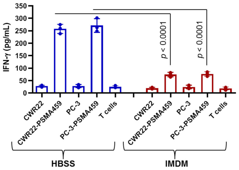
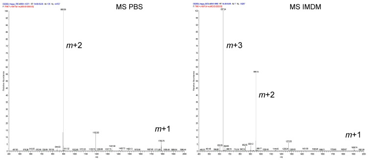
Prostate cancer poses an ongoing problem in the western world accounting for significant morbidity and mortality in the male population. Current therapy options are effective in treating most prostate cancer patients, but a significant number of patients progress beyond a manageable disease. For these patients, immunotherapy has emerged as a real option in the treatment of the late-stage metastatic disease. Unfortunately, even the most successful immunotherapy strategies have only led to a four-month increase in survival. One issue responsible for the shortcomings in cancer immunotherapy is the inability to stimulate helper CD4+ T cells via the HLA class II pathway to generate a potent antitumor response. Obstacles to proper HLA class II stimulation in prostate cancer vaccine design include the lack of detectable class II proteins in prostate tumors and the absence of defined class II specific prostate tumor antigens. Here, for the first time, we show that the insertion of a lysosomal thiol reductase (GILT) into prostate cancer cells directly enhances HLA class II antigen processing and results in increased CD4+ T cell activation by prostate cancer cells. We also show that GILT insertion does not alter the expression of prostate-specific membrane antigen (PSMA), an important target in prostate cancer vaccine strategies. Our study suggests that GILT expression enhances the presentation of the immunodominant PSMA459 epitope via the HLA class II pathway. Biochemical analysis showed that the PSMA459 peptide was cysteinylated under a normal physiologic concentration of cystine, and this cysteinylated form of PSMA459 inhibited T cell activation. Taken together, these results suggest that GILT has the potential to increase HLA class II Ag presentation and CD4+ T cell recognition of prostate cancer cells, and GILT-expressing prostate cancer cells could be used in designing cell therapy and/or vaccines against prostate cancer.
Keywords: CD4+ T cells; GILT; HLA class II; antigen presentation; cysteinylation; prostate cancer; prostate-specific membrane protein.
Conflict of interest statement
The authors have no financial conflict of interest.
Figures


Similar articles
-
Gamma-IFN-inducible-lysosomal thiol reductase modulates acidic proteases and HLA class II antigen processing in melanoma.Cancer Immunol Immunother. 2008 Oct;57(10):1461-70. doi: 10.1007/s00262-008-0483-8. Epub 2008 Mar 15. Cancer Immunol Immunother. 2008. PMID: 18343923 Free PMC article.
-
Prostate Cancer Immunotherapy: Exploiting the HLA Class II Pathway in Vaccine Design.J Clin Cell Immunol. 2015 Aug;6(4):351. doi: 10.4172/2155-9899.1000351. Epub 2015 Aug 26. J Clin Cell Immunol. 2015. PMID: 26807308 Free PMC article.
-
Identification of MHC class II-restricted T-cell epitopes in prostate-specific membrane antigen.Clin Cancer Res. 2003 Aug 15;9(9):3260-71. Clin Cancer Res. 2003. PMID: 12960111
-
Diverse cellular and organismal functions of the lysosomal thiol reductase GILT.Mol Immunol. 2015 Dec;68(2 Pt A):124-8. doi: 10.1016/j.molimm.2015.06.008. Epub 2015 Jun 23. Mol Immunol. 2015. PMID: 26116226 Free PMC article. Review.
-
HLA-Class II Artificial Antigen Presenting Cells in CD4+ T Cell-Based Immunotherapy.Front Immunol. 2019 May 17;10:1081. doi: 10.3389/fimmu.2019.01081. eCollection 2019. Front Immunol. 2019. PMID: 31156634 Free PMC article. Review.
Cited by
-
Therapeutic cancer vaccines: advancements, challenges, and prospects.Signal Transduct Target Ther. 2023 Dec 13;8(1):450. doi: 10.1038/s41392-023-01674-3. Signal Transduct Target Ther. 2023. PMID: 38086815 Free PMC article. Review.
-
Lower urinary dysfunction as a long-term effect of childhood vincristine treatment, with potential influences by sex and dose.Sci Rep. 2024 Jul 1;14(1):15049. doi: 10.1038/s41598-024-65313-9. Sci Rep. 2024. PMID: 38951167 Free PMC article.
-
Advances in Therapeutic Cancer Vaccines, Their Obstacles, and Prospects Toward Tumor Immunotherapy.Mol Biotechnol. 2025 Apr;67(4):1336-1366. doi: 10.1007/s12033-024-01144-3. Epub 2024 Apr 16. Mol Biotechnol. 2025. PMID: 38625508 Review.
References
-
- ACS . Cancer Facts & Figures 2010. American Cancer Society; Atlanta, GA, USA: 2010. p. 4.
MeSH terms
Substances
Grants and funding
LinkOut - more resources
Full Text Sources
Medical
Molecular Biology Databases
Research Materials
Miscellaneous
