. 2022 Dec 5;23(23):15324.
doi: 10.3390/ijms232315324.

Identification of Agents That Ameliorate Hyperphosphatemia-Suppressed Myogenin Expression Involved in the Nrf2/p62 Pathway in C2C12 Skeletal Muscle Cells

Affiliations

Identification of Agents That Ameliorate Hyperphosphatemia-Suppressed Myogenin Expression Involved in the Nrf2/p62 Pathway in C2C12 Skeletal Muscle Cells

Shu-Man Hsieh Li et al. Int J Mol Sci. .

Abstract

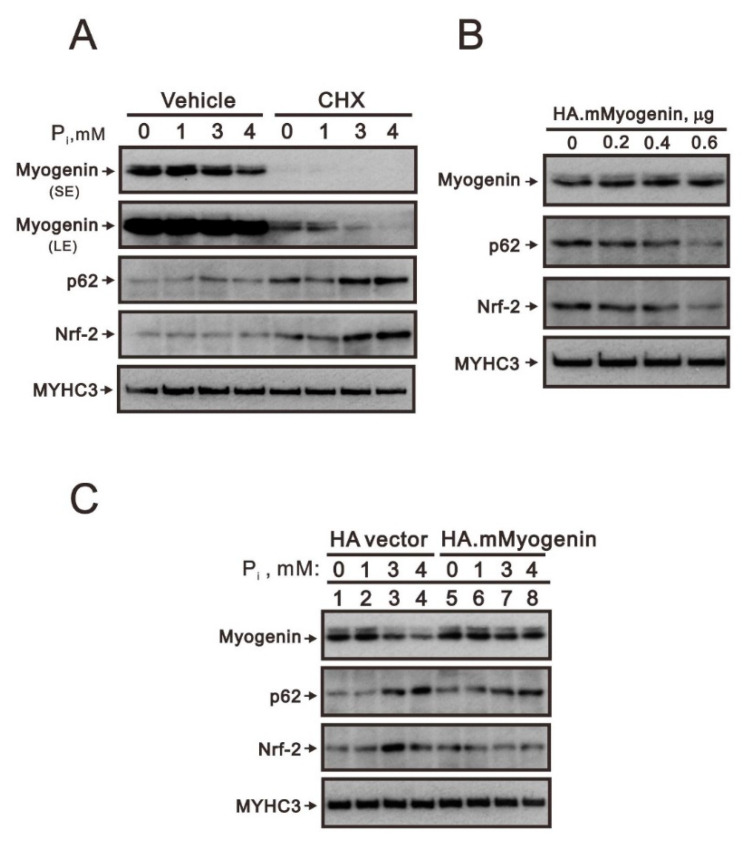
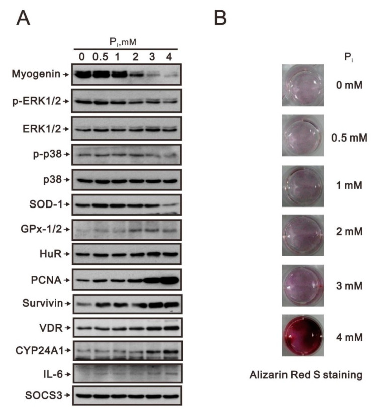
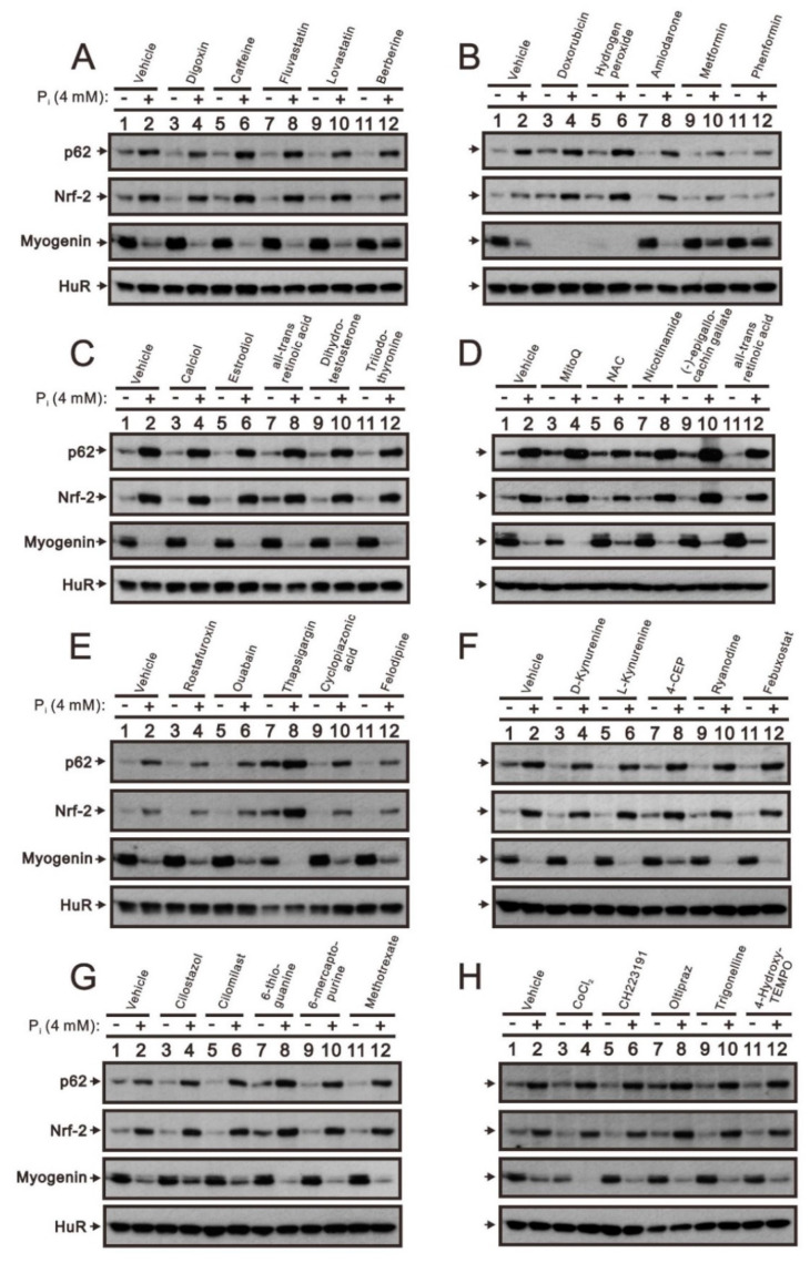
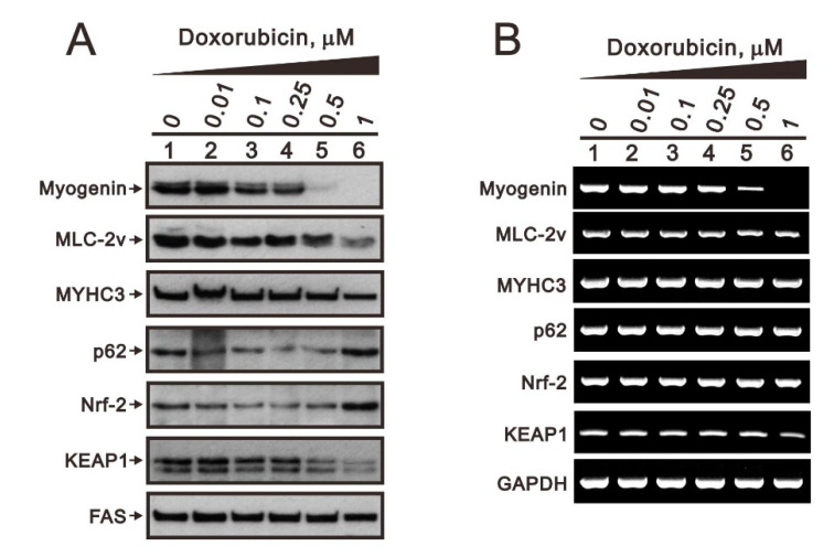
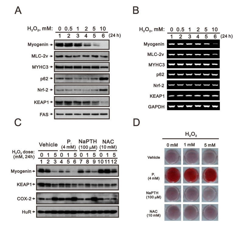
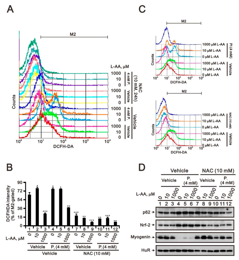
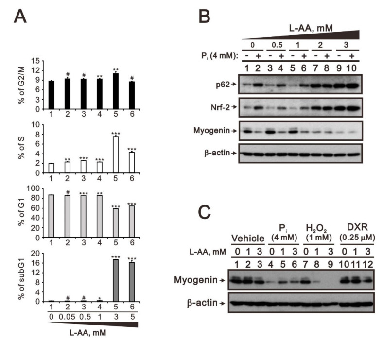
Hyperphosphatemia can occur as a result of reduced phosphate (Pi) excretion in cases of kidney dysfunction, which can induce muscle wasting and suppress myogenic differentiation. Higher Pi suppresses myogenic differentiation and promotes muscle atrophy through canonical (oxidative stress-mediated) and noncanonical (p62-mediated) activation of nuclear factor erythroid 2-related factor 2 (Nrf2) signaling. However, the crosstalk between myogenin and Nrf2/p62 and potential drug(s) for the regulation of myogenin expression needed to be addressed. In this study, we further identified that myogenin may negatively regulate Nrf2 and p62 protein levels in the mouse C2C12 muscle cell line. In the drug screening analysis, we identified N-acetylcysteine, metformin, phenformin, berberine, 4-chloro-3-ethylphenol, cilostazol, and cilomilast as ameliorating the induction of Nrf2 and p62 expression and reduction in myogenin expression that occur due to high Pi. We further elucidated that doxorubicin and hydrogen peroxide reduced the amount of myogenin protein mediated through the Kelch-like ECH-associated protein 1/Nrf2 pathway, differently from the mechanism of high Pi. The dual functional roles of L-ascorbic acid (L-AA) were found to be dependent on the working concentration, where concentrations below 1 mM L-AA reversed the effect of high Pi on myogenin and those above 1 mM L-AA had a similar effect of high Pi on myogenin when used alone. L-AA exacerbated the effect of hydrogen peroxide on myogenin protein and had no further effect of doxorubicin on myogenin protein. In summary, our results further our understanding of the crosstalk between myogenin and Nrf2, with the identification and verification of several potential drugs that can be applied in rescuing the decline of myogenin due to high Pi in muscle cells.

Keywords: L-ascorbic acid; hyperphosphatemia; myogenin; nuclear factor erythroid 2-related factor 2; p62.


Conflict of interest statement

The authors declare no conflict of interest.

Figures

Figure 1
Figure 1
The effects of myogenin induced by high Pi on Nrf2 and p62 proteins in differentiated C2C12 cells. (A) After induction of differentiation by culturing C2C12 cells in differentiation medium for 3 days, differentiated C2C12 cells were pretreated for 2 h with CHX (5 μg/mL) and then were treated for 24 h with the indicated concentrations of Pi. (B) Differentiated C2C12 cells were transiently transfected with indicated amounts of pSG5.HA. Myogenin for 4 h and further cultured for 20 h. (C) Differentiated C2C12 cells were transiently transfected with indicated amounts of pSG5.HA.Myogenin for 4 h and then treated with the indicated concentrations of Pi for 20 h. (AC) Immunoblot analysis of myogenin, p62, Nrf2, and MYHC3 (myosin heavy chain isoform 3) levels. MYHC3 was a protein loading control. SE: shorter exposure; LE: longer exposure.
Figure 2
Figure 2
The effects of high Pi on myogenin and its associated proteins in differentiated C2C12 cells. After induction of differentiation by culturing C2C12 cells in differentiation medium for 3 days, differentiated C2C12 cells were treated with the indicated concentration of Pi for 24 h. (A) Cell lysates were subject to immunoblot analysis against indicated proteins. (B) Cells treated with Pi were stained with Alizarin Red S dye.
Figure 3
Figure 3
The drug screening platform for the rescue effect on the high Pi in differentiated C2C12 cells. After induction of differentiation by culturing C2C12 cells in differentiation medium for 3 days, differentiated C2C12 cells were treated with the indicated drug (A): vehicle, digoxin, caffeine, fluvastatin, lovastatin, and berberine; (B): vehicle, doxorubicin, hydrogen peroxide, amiodarone, metformin, and phenformin; (C): vehicle, calciol, estradiol, all-trans retinoic acid, dihydrotestosterone, and triiodothyronine; (D): vehicle, MitoQ, NAC, nicotinamide, (-)-epigallocachin gallate, and all-trans retinoic acid; (E): vehicle, rostafuroxin, ouabain, thapsigargin, cyclopiazonic acid, and felodipine; (F): vehicle, D-kynurenine, L-kynurenine, 4-CEP, ryanodine, and febuxostat; (G): vehicle, cilostazol, cilomilast, 6-thioguanine, 6-mercaptopurine, and methotrexate; (H): vehicle, CoCl2, CH223191, oltipraz, trigonelline, and 4-hydroxy-TEMPO) with 4 mM Pi for 24 h. Cell lysates were subject to immunoblot analysis against p62, Nrf2, and myogenin proteins. HuR was a protein loading control.
Figure 4
Figure 4
The effects of doxorubicin on the Keap1/Nrf2/p21/myogenin pathway in differentiated C2C12 cells. After induction of differentiation by culturing C2C12 cells in differentiation medium for 3 days, differentiated C2C12 cells were treated with the indicated concentration of doxorubicin for 20 h. (A) Cell lysates were subject to immunoblot analysis against myogenin, MLC-2v, MYHC3, p62, Nrf2, and Keap1 proteins. FAS was used as loading control. (B) Effects of doxorubicin on the mRNA expression of myogenin, MLC-2v, MYHC3, p62, Nrf2, and Keap1 determined by RT-PCR. GAPDH was used as loading control.
Figure 5
Figure 5
The effects of hydrogen peroxide on the Keap1/Nrf2/p62/myogenin pathway in differentiated C2C12 cells. (A,B) After induction of differentiation by culturing C2C12 cells in differentiation medium for 3 days, differentiated C2C12 cells were treated with the indicated concentration of hydrogen peroxide (H2O2) for 24 h. Cell lysates were subject to immunoblot analysis against indicated proteins. (A) Cell lysates were subject to immunoblot analysis against myogenin, MLC-2v, MYHC3, p62, Nrf2, and Keap1 proteins. FAS was used as loading control. (B) Effects of doxorubicin on the mRNA expression of myogenin, MLC-2v, MYHC3, p62, Nrf2, and Keap1 determined by RT-PCR. GAPDH was used as loading control. (C,D) Differentiated C2C12 cells were treated with the indicated concentration of hydrogen peroxide with 4 mM Pi, 100 μM NaPTH, or 10 mM NAC for 24 h. (C) Cell lysates were subject to immunoblot analysis against myogenin, Keap1, and Cox-2 proteins. HuR was a protein loading control. (D) Cells treated with indicated drugs were stained with Alizarin Red S dye.
Figure 6
Figure 6
The effects of L-ascorbic acid on high Pi-induced ROS generation in differentiated C2C12 cells. (A) Differentiated C2C12 cells were treated with the indicated concentrations of L-ascorbic acid (L-AA) combined with 5 mM NAC or 4 mM Pi for 24 h. (A,B) Cytosolic ROS levels were determined based on H2DCFDA fluorescence using the FL1-H channel: the x-axis represents H2DCFDA intensity; the y-axis indicates the cell counts at the corresponding fluorescence intensity. Bars depict the mean ± SD. * p < 0.05; ** p < 0.01; *** p < 0.001. (C) Bar graph summarizing the data from panel A as percentages of cells with increased ROS production (% M2 gated). (D) Cell lysates were subject to immunoblot analysis against p62, Nrf2, and myogenin proteins. HuR was used as loading control.
Figure 7
Figure 7
The effects of L-AA on the cell cycle profile and the crosstalk with Pi, doxorubicin, and hydrogen peroxide in differentiated C2C12 cells. (A) Differentiated C2C12 cells were treated with the indicated concentrations of L-AA for 24 h. The cell cycle profile was analyzed with PI dye. Data are presented as a percentage of the control. Bars depict the mean ± SD. # p > 0.05; * p < 0.05; ** p < 0.01; *** p < 0.001. (B) Differentiated C2C12 cells were treated with the indicated concentrations of L-AA combined with 4 mM Pi for 24 h. Cell lysates were subject to immunoblot analysis against p62, Nrf2, and myogenin proteins. β-actin was used as loading control. (C) Differentiated C2C12 cells were treated with the indicated concentrations of L-AA combined with 4 mM Pi, 1mM hydrogen peroxide (H2O2), or 0.25 µM doxorubicin (DXR) for 24 h. Cell lysates were subject to immunoblot analysis against myogenin protein. β-actin was used as loading control.

Similar articles

References

    1. Hruska K.A., Mathew S., Lund R., Qiu P., Pratt R. Hyperphosphatemia of chronic kidney disease. Kidney Int. 2008;74:148–157. doi: 10.1038/ki.2008.130. - DOI - PMC - PubMed
    1. Lederer E. Regulation of serum phosphate. J. Physiol. 2014;592:3985–3995. doi: 10.1113/jphysiol.2014.273979. - DOI - PMC - PubMed
    1. Boengler K., Kosiol M., Mayr M., Schulz R., Rohrbach S. Mitochondria and ageing: Role in heart, skeletal muscle and adipose tissue. J. Cachexia Sarcopenia Muscle. 2017;8:349–369. doi: 10.1002/jcsm.12178. - DOI - PMC - PubMed
    1. Deane C.S., Ely I.A., Wilkinson D.J., Smith K., Phillips B.E., Atherton P.J. Dietary protein, exercise, ageing and physical inactivity: Interactive influences on skeletal muscle proteostasis. Proc. Nutr. Soc. 2021;80:106–117. doi: 10.1017/S0029665120007879. - DOI - PubMed
    1. Chung L.H., Liu S.T., Huang S.M., Salter D.M., Lee H.S., Hsu Y.J. High phosphate induces skeletal muscle atrophy and suppresses myogenic differentiation by increasing oxidative stress and activating Nrf2 signaling. Aging. 2020;12:21446–21468. doi: 10.18632/aging.103896. - DOI - PMC - PubMed

MeSH terms