. 2022 Dec 5;23(23):15328.
doi: 10.3390/ijms232315328.

Osteopontin Splicing Isoforms Contribute to Endometriotic Proliferation, Migration, and Epithelial-Mesenchymal Transition in Endometrial Epithelial Cells

Affiliations

Osteopontin Splicing Isoforms Contribute to Endometriotic Proliferation, Migration, and Epithelial-Mesenchymal Transition in Endometrial Epithelial Cells

Nguyen-Tuong Ho et al. Int J Mol Sci. .

Abstract

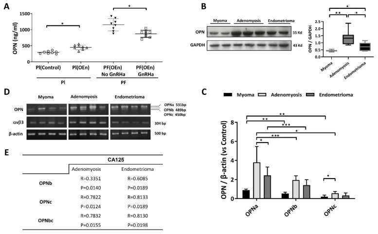
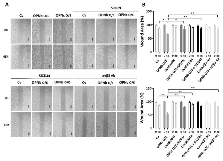
Osteopontin (OPN) isoforms, including OPNb and OPNc, promote malignancy and may contribute to the pathogenesis of endometriosis, a benign disorder with multiple characteristics resembling malignant tumors. In our experiments, OPNb and OPNc were significantly overexpressed in both endometriosis and adenomyosis compared to the normal endometrium. Upregulation of CD44v and the epithelial-mesenchymal transition (EMT) process was also present in endometriotic lesions. Overexpression of OPNb and OPNc splicing variants in endometriotic cells evoked morphological changes, actin remodeling, cell proliferation, cell migration, and EMT through binding OPN ligand receptors CD44 and αvβ3, subsequently activating the PI3K and NF-ĸB pathways. We elucidated the causal role of OPN splice variants in regulating endometriotic cell growth, which may promote the development of OPN-targeted therapies for patients suffering from endometriotic disorders.

Keywords: CD44; endometriosis; epithelial-mesenchymal transition; migration; osteopontin splicing variants; proliferation; αvβ3.


Conflict of interest statement

The authors declare no conflict of interest.

Figures

Figure 1
Figure 1
Expression of OPN and OPN isoforms in endometriotic lesions. (A) OPN levels in plasmas from women with myomas (n = 8, control) and women with ovarian endometrioma (n = 8), in peritoneal fluids from women with ovarian endometrioma (n = 8) before and after GnRH agonist treatment. (B) Representative blot image and quantitative analysis of Western blot data on OPN in myoma (n = 12), adenomyosis (n = 12), and ovarian endometrioma tissues (n = 12). The normalized OPN expression in adenomyosis was the highest, followed by the endometriosis tissues compared to the myoma tissues, p < 0.05. (C,D) Expressions of OPN isoforms (OPNa, OPNb, OPNc) in myoma, adenomyosis, and ovarian endometriosis tissues. All OPN isoforms were upregulated in endometriotic lesions, especially in adenomyotic tissue (p < 0.05). All OPN isoforms were expressed at significantly higher levels in endometriotic lesions than in the myoma control group (p < 0.05). (E) Linear correlation between CA125 and OPN isoforms in different endometriotic lesions. Plots represent the mean ± standard error of the mean (SEM). Pl: plasma; OEn: Ovarian endometrioma(s); PF: peritoneal fluid; GnRHa: gonadotropin-releasing hormone agonist; OPN: osteopontin; CA125: cancer antigen 125. (*) indicates a significant difference with p < 0.05. (**) indicates a significant difference with p < 0.01. (***) indicates a significant difference with p < 0.001.
Figure 2
Figure 2
Differential expression of OPN ligand-receptor CD44s and its variant CD44v in endometriotic samples. (A) Quantification of proteomic data from controls and endometriotic tissues showed increased CD44v expression in endometriosis tissues. In contrast, both adenomyosis and ovarian endometriosis demonstrated slightly reduced CD44s expression. (B) The representative electrophoretogram of CD44s, CD44v, and β-actin in myoma, adenomyosis, and endometrioma is shown. (C) Normalized CD44 mRNA expression demonstrated the overexpression of CD44v in adenomyosis and ovarian endometrioma samples. In comparison to myoma cells, the CD44s variant was found to be overexpressed in adenomyosis cells but was diminished in endometrioma cells. (D) The linear correlation between the expression levels of CD44 variants and OPN isoforms in various endometriotic tissues. Plots represent the mean ± SEM. CD44s: CD44 standard; CD44v: CD44 variant. (*) indicates a significant difference with p < 0.05. (**) indicates a significant difference with p < 0.01. (***) indicates a significant difference with p < 0.001.
Figure 3
Figure 3
EMT markers and cell adhesion molecules (CAM) in myoma, adenomyosis and ectopic endometriosis lesions. (A) Representative chemiluminescence image of Western blot data demonstrated increased EMT markers in endometriosis. E-cadherin was underexpressed in adenomyotic lesions. N-cadherin and Vimentin were both found to be overexpressed in myoma tissue and endometriotic lesions. Additionally, the Vimentin concentration was extremely high in adenomyosis compared with endometrioma, myoma, and normal endometrium (p < 0.05). (B) The mRNA expression levels of EMT markers, including N-cadherin, E-cadherin, Vimentin, and CAM markers, ICAM and VCAM, were detected by quantitative real-time PCR, as shown in the electrophoretogram. (C) Analysis and normalization of the mRNA levels of EMT and CAM markers. E-cadherin was downregulated, while N-cadherin, Vimentin, ICAM and VCAM expression was upregulated in endometriotic lesions (p < 0.05). Plots represent the mean ± SEM. Ecad: E-cadherin; Ncad: N-cadherin; ICAM: intercellular adhesion molecule; VCAM: vascular cell adhesion molecule. (*) indicates a significant difference with p < 0.05. (**) indicates a significant difference with p < 0.01. (***) indicates a significant difference with p < 0.001.
Figure 4
Figure 4
The efficiency of OPN isoform transfection and CD44 knockdown in OPN-overexpressing HEC1A cells was identified by real-time PCR. (A) Schematic illustration of OPN isoforms OPNa, OPNb, and OPNc. (B) OPN splice variants, OPNb and OPNc, were transfected into endometriotic cells. The vector only was used as a control (Cv). (C) The expression of OPN splice variants in the OPNb- or OPNc-transfected cell lines was identified by quantitative real-time PCR. (D) OPN silencing with OPN-siRNA in OPNb/OPNc-overexpressing endometriotic cells. (E) CD44 knockdown efficiency was identified by real-time PCR. Cv: control vector; O/E: overexpression. Plots represent the mean ± SEM, n = 4. (*) indicates a significant difference with p < 0.05. (**) indicates a significant difference with p < 0.01. (***) indicates a significant difference with p < 0.001.
Figure 5
Figure 5
The effect of OPN on actin filament remodeling of HEC1A cells and the expression of OPN ligand receptors or EMT markers. (A) Phalloidin conjugates to the orange fluorescent dye phalloidin-tetramethylrhodamine (TRITC) for actin labeling and the blue fluorescent dye (DAPI) for nuclei and was observed under a Leica TCS SP5 confocal microscope. Overexpression of OPN splice variants induced pseudopodia extension. In contrast, the knockdown of OPN simultaneously reduced actin polymerization. The knockdown of CD44 or inhibition of αVβ3 integrin activity with the corresponding antibody concurrently reduces actin filament remodeling. (B) The PCR products of OPN ligand receptors (αvβ3, CD44s, and CD44v) and EMT markers (N-cad, E-cad, and Vimentin) in OPN-overexpressing (O/E) cells were observed using an electrophoretogram. (C) The mRNA expression levels of CD44v, CD44s, Vimentin, N-cadherin, and E-cadherin were detected by quantitative real-time PCR. Higher expression levels of CD44s and CD44v were found in OPN O/E cells. Additionally, the expression of CD44s in the OPNb-overexpressing cells was significantly higher than that in the other groups. Enhanced Vimentin and N-cadherin mRNA levels were found in conjunction with a reduced E-cadherin level in OPN O/E cells. Plots represent the mean ± SEM, n = 4. CD44s: CD44 standard; CD44v: CD44 variant; O/E: overexpression; si: siRNA; αVβ3 Ab: anti-αVβ3 integrin antibody; Ecad: E-cadherin; Ncad: N-cadherin. (*) indicates a significant difference with p < 0.05. (**) indicates a significant difference with p < 0.01. (***) indicates a significant difference with p < 0.001.
Figure 6
Figure 6
The effect of OPN isoforms on HEC1A cell migration. (A) Both OPNb and OPNc promoted cellular migratory ability, but the latter showed a significantly higher stimulation of endometriotic cell migration. Knockdown OPN or suppression of OPN ligand receptors (αvβ3 and CD44) reversed the changes in the wound healing area. (B) Plots show the differences in migration between control cells and OPN-overexpressing cells before and after suppression. Plots represent the mean ± SEM, n = 4. Cv: control vector, O/E: overexpression, si: siRNA, αVβ3 Ab: anti-αVβ3 integrin antibody. (*) indicates a significant difference with p < 0.05. (**) indicates a significant difference with p < 0.01.
Figure 7
Figure 7
The effect of OPN on cell proliferation in OPN-overexpressing HEC1A cells. (A) The OPNb- or OPNc-overexpressing cells showed enhanced proliferation. The morphological change into a slender phenotype similar to the mesenchymal type was also found in the OPN-overexpressing cells. In contrast, through the depletion of OPN and OPN ligand receptors (CD44 and αvβ3), the cell phenotypes resembled those of the control cells. (B) The impact of OPN variants on cell proliferation was determined by using the BrdU Cell Proliferation Assay Kit. The results also showed that proliferation was more prominent in OPNb-overexpressing cells than in OPNc-overexpressing and control cells. Plots represent the mean ± SEM, n = 4. Cv: control vector; O/E: overexpression; si: siRNA, αVβ3 Ab: anti-αVβ3 integrin antibody. (*) indicates a significant difference with p < 0.05. (**) indicates a significant difference with p < 0.01. (***) indicates a significant difference with p < 0.001.
Figure 8
Figure 8
The effect of OPN variants on HEC1A cell migration via the PI3K or NF-κB signaling pathway. (A,B) A scratch wound assay was performed to investigate endometriotic cell migration. OPN isoforms can lead to cell migration, but treatment with 10 μM wortmannin (PI-3K inhibitor) or 10 μM CAPE (NF-κB inhibitor) for 24 h can slow down or suppress cell migration. (C) Wortmannin and CAPE inhibited proliferation in cells overexpressing OPN isoforms. (D) Nuclear translocation of NF-κB was inhibited by OPN suppression. Plots represent the mean ± SEM, n = 4. (*) indicates a significant difference with p < 0.05. (**) indicates a significant difference with p < 0.01. (***) indicates a significant difference with p < 0.001.
Figure 9
Figure 9
Schematic illustration of the role of OPN isoforms in endometriosis pathogenesis. OPNb and OPNc bind to OPN-ligand receptors CD44 and αvβ3 integrin, activating the PI3K and NFkB pathways, thereby inducing endometriotic cellular migration, proliferation, and the epithelial-mesenchymal transition process.

Similar articles

Cited by

References

    1. Bulletti C., Coccia M.E., Battistoni S., Borini A. Endometriosis and infertility. J. Assist. Reprod. Genet. 2010;27:441–447. doi: 10.1007/s10815-010-9436-1. - DOI - PMC - PubMed
    1. Lamouille S., Xu J., Derynck R. Molecular mechanisms of epithelial-mesenchymal transition. Nat. Rev. Mol. Cell Biol. 2014;15:178–196. doi: 10.1038/nrm3758. - DOI - PMC - PubMed
    1. Loh C.-Y., Chai J.Y., Tang T.F., Wong W.F., Sethi G., Shanmugam M.K., Chong P.P., Looi C.Y. The E-Cadherin and N-Cadherin Switch in Epithelial-to-Mesenchymal Transition: Signaling, Therapeutic Implications, and Challenges. Cells. 2019;8:1118. doi: 10.3390/cells8101118. - DOI - PMC - PubMed
    1. Liu C.-Y., Lin H.-H., Tang M.-J., Wang Y.-K. Vimentin contributes to epithelial-mesenchymal transition cancer cell mechanics by mediating cytoskeletal organization and focal adhesion maturation. Oncotarget. 2015;6:15966–15983. doi: 10.18632/oncotarget.3862. - DOI - PMC - PubMed
    1. Xiong Y., Liu Y., Xiong W., Zhang L., Liu H., Du Y., Li N. Hypoxia-inducible factor 1alpha-induced epithelial-mesenchymal transition of endometrial epithelial cells may contribute to the development of endometriosis. Hum. Reprod. 2016;31:1327–1338. doi: 10.1093/humrep/dew081. - DOI - PubMed