. 2022 Nov 23;27(23):8144.
doi: 10.3390/molecules27238144.

Targeted Nanoparticles for the Binding of Injured Vascular Endothelium after Percutaneous Coronary Intervention

Affiliations

Targeted Nanoparticles for the Binding of Injured Vascular Endothelium after Percutaneous Coronary Intervention

Pennapa Mungchan et al. Molecules. .

Abstract

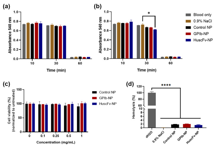
Percutaneous coronary intervention (PCI) is a common procedure for the management of coronary artery obstruction. However, it usually causes vascular wall injury leading to restenosis that limits the long-term success of the PCI endeavor. The ultimate objective of this study was to develop the targeting nanoparticles (NPs) that were destined for the injured subendothelium and attract endothelial progenitor cells (EPCs) to the damaged location for endothelium regeneration. Biodegradable poly(lactic-co-glycolic acid) (PLGA) NPs were conjugated with double targeting moieties, which are glycoprotein Ib alpha chain (GPIbα) and human single-chain antibody variable fragment (HuscFv) specific to the cluster of differentiation 34 (CD34). GPIb is a platelet receptor that interacts with the von Willebrand factor (vWF), highly deposited on the damaged subendothelial surface, while CD34 is a surface marker of EPCs. A candidate anti-CD34 HuscFv was successfully constructed using a phage display biopanning technique. The HuscFv could be purified and showed binding affinity to the CD34-positive cells. The GPIb-conjugated NPs (GPIb-NPs) could target vWF and prevent platelet adherence to vWF in vitro. Furthermore, the HuscFv-conjugated NPs (HuscFv-NPs) could capture CD34-positive cells. The bispecific NPs have high potential to locate at the damaged subendothelial surface and capture EPCs for accelerating the vessel repair.

Keywords: biodegradable nanoparticles; endothelium regeneration; percutaneous coronary intervention; single-chain antibody variable fragment; vascular injury.


Conflict of interest statement

The authors declare no conflict of interest.

Figures

Figure 1
Figure 1
Characterization and binding tests of soluble anti-CD34 HuscFv. (a) The HuscFv (arrowhead) produced by the huscfv-positive Rosetta E. coli clone as determined by Western blotting. Lane M, Pre-stained standard protein marker; Lanes 1–5, HuscFv of randomly picked transformed E. coli colonies. (b) Binding test of crude HuscFv from selected phage-transformed HB2151 E. coli clone to recombinant CD34 by indirect ELISA. BSA served as negative control antigen. HB2151, lysate of original HB2151 E. coli as background binding control; Positive control, mouse anti-CD34 Ab; Dotted blackline, the cut-off absorbance between positive and negative ELISA that determined from the highest absorbance of HB2151 E. coli control. Presented data are mean ± SD (n = 3). Representative of binding results of purified HuscFv to (c) HUVECs and (d) KG-1a cells by flow cytometry analysis.
Figure 2
Figure 2
Morphology and distribution of NPs via FE-SEM. (a) Control NPs and (b) conjugated NPs examined by FE-SEM. Scale bar represents 500 nm.
Figure 3
Figure 3
Cytocompatibility and hemocompatibility of NPs. Absorbance 540 nm of whole blood samples after treatment with PLGA NP samples for 10, 30 and 60 min at the NP concentrations of (a) 0.5 mg/mL and (b) 1.0 mg/mL. Normal saline (0.9% NaCl) served as negative control. (c) Cytotoxicity of HUVECs exposed to various concentrations of NP preparations for 24 h by MTS assay. (d) Hemolysis test of whole blood samples treated with various NP preparations. The absorbance of 545 nm for determining hemolysis when treated anticoagulated blood with NP preparations for 2 h at the NP concentration of 1.0 mg/mL. The anticoagulated blood treated with 0.9% NaCl and dH2O served as negative and positive hemolysis control, respectively. Blank, uncoated NPs served as control NPs. Data are presented as mean ± SD (n = 3). * p < 0.05 and **** p < 0.0001.
Figure 4
Figure 4
Binding tests of HuscFv-NPs to (a) HUVECs and (b) KG-1a cells by flow cytometry. The percentage of RhB-positive cells of (c) HUVECs and (d) KG-1a cells, and mean fluorescence intensity (MFI) of conjugated-NPs binding to (e) HUVECs and (f) KG-1a cells. Positive and negative controls were anti-CD34 Ab-conjugated NPs (anti-CD34-NP) and RhB-loaded NPs (RhB NP), respectively. Data are presented as mean ± SD (n = 3). ** p < 0.01, *** p < 0.001 and **** p < 0.0001.
Figure 5
Figure 5
In vitro targeting efficiency of GPIb-NPs to vWF and prevention of platelet adherence to vWF-coated surface. (a) Fluorescence intensity (a.u.) of GPIb-NPs adhering on vWF-coated surfaces. Data are presented as mean ± SD (n = 6). (b) Binding competition of GPIb-NPs to vWF-coated surface in the presence of free vWF as determined by fluorescence intensity (a.u.). C6-loaded NPs (C6 NP) served as control NPs. (c) The percentage of platelet adherence to vWF-coated surface in the presence of different NP samples quantified by LDH assays, which was determined at absorbance at 490 nm, then normalized to the PRP-only group. Platelet-poor-plasma (PPP) and PRP only served as negative and positive controls, respectively. Data are presented as mean ± SD (n = 3). * p < 0.05 and **** p < 0.0001.

References

    1. Malakar A.K., Choudhury D., Halder B., Paul P., Uddin A., Chakraborty S. A review on coronary artery disease, its risk factors, and therapeutics. J. Cell Physiol. 2019;234:16812–16823. doi: 10.1002/jcp.28350. - DOI - PubMed
    1. Alvarez-Alvarez M.M., Zanetti D., Carreras-Torres R., Moral P., Athanasiadis G. A survey of sub-Saharan gene flow into the Mediterranean at risk loci for coronary artery disease. Eur. J. Hum. Genet. 2017;25:472–476. doi: 10.1038/ejhg.2016.200. - DOI - PMC - PubMed
    1. World Health Organization Cardiovascular Diseases (CVDs) [(accessed on 8 May 2022)]. Available online: https://www.who.int/en/news-room/fact-sheets/detail/cardiovascular-disea...
    1. Khan S.Q., Ludman P.F. Percutaneous coronary intervention. Medicine. 2022;50:437–444. doi: 10.1016/j.mpmed.2022.04.008. - DOI
    1. Cassar A., Holmes D.R., Jr., Rihal C.S., Gersh B.J. Chronic Coronary Artery Disease: Diagnosis and Management. Mayo. Clin. Proc. 2009;84:1130–1146. doi: 10.4065/mcp.2009.0391. - DOI - PMC - PubMed

LinkOut - more resources