. 2022 Dec 6;27(23):8623.
doi: 10.3390/molecules27238623.

FOXO3a Mediates Homologous Recombination Repair (HRR) via Transcriptional Activation of MRE11, BRCA1, BRIP1, and RAD50

Affiliations

FOXO3a Mediates Homologous Recombination Repair (HRR) via Transcriptional Activation of MRE11, BRCA1, BRIP1, and RAD50

Gozde Inci et al. Molecules. .

Abstract

To test whether homologous recombination repair (HRR) depends on FOXO3a, a cellular aging model of human dermal fibroblast (HDF) and tet-on flag-h-FOXO3a transgenic mice were studied. HDF cells transfected with over-expression of wt-h-FOXO3a increased the protein levels of MRE11, BRCA1, BRIP1, and RAD50, while knock-down with siFOXO3a decreased them. The protein levels of MRE11, BRCA1, BRIP1, RAD50, and RAD51 decreased during cellular aging. Chromatin immunoprecipitation (ChIP) assay was performed on FOXO3a binding accessibility to FOXO consensus sites in human MRE11, BRCA1, BRIP1, and RAD50 promoters; the results showed FOXO3a binding decreased during cellular aging. When the tet-on flag-h-FOXO3a mice were administered doxycycline orally, the protein and mRNA levels of flag-h-FOXO3a, MRE11, BRCA1, BRIP1, and RAD50 increased in a doxycycline-dose-dependent manner. In vitro HRR assays were performed by transfection with an HR vector and I-SceI vector. The mRNA levels of the recombined GFP increased after doxycycline treatment in MEF but not in wt-MEF, and increased in young HDF comparing to old HDF, indicating that FOXO3a activates HRR. Overall, these results demonstrate that MRE11, BRCA1, BRIP1, and RAD50 are transcriptional target genes for FOXO3a, and HRR activity is increased via transcriptional activation of MRE11, BRCA1, BRIP1, and RAD50 by FOXO3a.

Keywords: BRCA1; BRIP1; FOXO3a; MRE11; RAD50; homologous recombination repair.


Conflict of interest statement

The authors declare no conflict of interest.

Figures
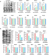
Figure 1
Figure 1
(A) The protein levels of MRE11, BRCA1, BRIP1, RAD50, RAD51, FOXO3a, NBS1, BARD1, PALB2, and β-actin were measured by Western blotting in HDF cells transfected with increasing amounts of wt-h-FOXO3a plasmid. HDF cells of PD24 (young) were transfected with 0, 300, 600, and 1200 ng of wt-h-FOXO3a using Lipofectamine 3000. The HDF cells were then cultured for 24 h. Transfection efficiency was determined by pCMV-beta-Gal-transfected HDF. Relative band intensities of MRE11, BRCA1, BRIP1, RAD50, RAD51, NBS1, BARD1, PALB2, and FOXO3a versus β-actin were measured by densitometry and the data were plotted as histograms. (B) The protein levels of MRE11, BRCA1, BRIP1, and RAD50 were measured in siFOXO3a-transfected HDF PD24 (young) cells by Western blotting. Standard deviations as error bars were obtained from three different experiments. Statistical significance is indicated as * p < 0.05, ** p < 0.01, and *** p < 0.001.
Figure 2
Figure 2
The protein levels of MRE11, BRCA1, BRIP1, RAD50, RAD51, BRCA2, FOXO3a, and β-actin were measured by Western blotting. Cell extracts were prepared from HDF cells at PD24 (“young”), PD30, PD36 (“middle”), PD46, and PD56 (“old”). Relative band intensities of MRE11, BRCA1, BRIP1, RAD50, RAD51, BRCA2, and FOXO3a versus β-actin were measured by densitometry and the data were plotted as histograms. Standard deviations as error bars were obtained from three different experiments. Statistical significance is indicated as * p < 0.05, ** p < 0.01, and *** p < 0.001.
Figure 3
Figure 3
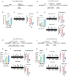
MRE11, BRCA1, BRIP1, and RAD50 promoter activities were measured in young HDF cells transfected with increasing levels of wt-h-FOXO3a plasmid. FOXO3a binding to FOXO consensus sites of each promoter was measured by ChIP assay during cellular aging of HDF. The schematic diagrams of (A) human MRE11 promoter–luciferase (pGL3-hMRE11), (D) human BRCA1 promoter–luciferase (pGL3-hBRCA1), (G) human BRIP1 promoter–luciferase (pGL3-hBRIP1), and (J) human RAD50 promoter–luciferase (pGL3-hRAD50) constructs depicting the FOXO-binding consensus sites and ChIP-primer-binding locations were shown. HDF cells were transfected with 0, 300, 600, and 1200 ng of wt-h-FOXO3a and (B) pGL3-hMRE11, (E) pGL3-hBRCA1, (H) pGL3-BRIP1, and (K) pGL3-RAD50 plasmids using Lipofectamine 3000. HDF cells were then incubated in DMEM for 24 h. The luciferase activity of the cell extracts was measured with a luminometer (GloMAX 20/20, Promega). Cell lysates were prepared from HDF cells of PD26 (“young”) and PD44 (“old”). Cell lysates were sonicated on ice to obtain sheared average DNA fragments of 300 bp to 1000 bp. Immunoprecipitation was carried out as described in the Materials and Methods section. Isolated genomic DNA from immunoprecipitation was used in the ChIP-PCR reaction. ChIP-PCR amplification of FOXO3a-binding consensus sites in (C) the human MRE11 promoter, (F) the human BRCA1 promoter, (I) the human BRIP1 promoter, and (L) the human RAD50 promoter was performed as described in the Materials and Methods section. The PCR products were separated by electrophoresis in 1.5% agarose gel. Standard deviations as error bars were obtained from three different experiments. Statistical significance is indicated as * p < 0.05, ** p < 0.01, and *** p < 0.001.
Figure 4
Figure 4
FOXO3a-dependent upregulation of MRE11, BRCA1, BRIP1, and RAD50 mRNA and protein expression in tet-on flag-h-FOXO3a transgenic mice. The tet-on flag-h-FOXO3a transgenic mice were used to test flag-h-FOXO3a-dependent gene expression in the tail-tips of the mice. Oral administration of 0.2 mL of doxycycline (0, 0.02, 0.2, and 2 mg in 1% sucrose) solution was performed on the transgenic mice once a day for 2 days. (A) RNAs were prepared from the tail-tips on day 3. RNA was analyzed by quantitative real-time RT-PCR as described in the Materials and Methods section. Relative amounts of flag-h-FOXO3a, MRE11, BRCA1, BRIP1, RAD50, and mouse FOXO3a mRNAs versus β-actin mRNA were measured and the data were plotted as histograms. (B) Cell extracts were prepared from the tail-tip samples on day 3. Protein levels were analyzed by Western blotting using anti-flag, anti-MRE11, anti-BRCA1, anti-BRIP1, anti-RAD50, anti-FOXO3a, and anti-β-actin antibodies. Relative band intensities of flag-h-FOXO3a, MRE11, BRCA1, BRIP1, RAD50, and mFOXO3a versus β-actin were measured by densitometry and the data were plotted as histograms. Standard deviations as error bars were obtained from three different experiments. Statistical significance is indicated as * p < 0.05, ** p < 0.01, and *** p < 0.001.
Figure 5
Figure 5
In vitro HRR activity was measured by transfection of MEF and wt-MEF with DR-GFP (Addgene, #26475) plasmid and I-SceI (Addgene, #26477) plasmid. Following transfection of the MEFs with the HRR vector and I-Scel vector, the MEF cells were again treated with three concentrations of doxycycline or control (0, 0.5, 1.5, and 4.5 μg/mL) and wt-MEF cells were treated with 4.5 μg/mL of doxycycline in DMEM-10% FBS for 48 h. HDFs were also transfected with the HRR vector and I-Scel vector and incubated for 48 h. Old HDF cells were transfected with wt-h-FOXO3a for 24 h to induce FOXO3a before HRR assay to test the restoration of HRR activity. Total RNAs were isolated and RT-qPCRs for recombined and unrecombined GFP were performed to measure HRR activity. (A) Schematic diagram depicting the in vitro HRR assay. (B) RT-qPCR for total RNA was performed to detect recombined and unrecombined GFP in MEF (from tet-on h-FOXO3a transgenic mice) and (C) wt-MEF (from wild-type mice). Statistical significance is indicated as *** p < 0.001, ns: not significant.
Figure 6
Figure 6
A schematic diagram showing up-regulation of HRR activity via transcriptional activation of target HRR genes by FOXO3a.

Similar articles

References

    1. Lieber M.R. The Mechanism of Human Nonhomologous DNA End Joining. J. Biol. Chem. 2008;283:1–5. doi: 10.1074/jbc.R700039200. - DOI - PubMed
    1. Johnson R.D. Sister chromatid gene conversion is a prominent double-strand break repair pathway in mammalian cells. EMBO J. 2000;19:3398–3407. doi: 10.1093/emboj/19.13.3398. - DOI - PMC - PubMed
    1. Krejci L., Altmannova V., Spirek M., Zhao X. Homologous recombination and its regulation. Nucleic Acids Res. 2012;40:5795–5818. doi: 10.1093/nar/gks270. - DOI - PMC - PubMed
    1. Lieber M.R. The Mechanism of Double-Strand DNA Break Repair by the Nonhomologous DNA End-Joining Pathway. Annu. Rev. Biochem. 2010;79:181–211. doi: 10.1146/annurev.biochem.052308.093131. - DOI - PMC - PubMed
    1. Wyman C., Ristic D., Kanaar R. Homologous recombination-mediated double-strand break repair. DNA Repair. 2004;3:827–833. doi: 10.1016/j.dnarep.2004.03.037. - DOI - PubMed