. 2022 Nov 22;12(23):4110.
doi: 10.3390/nano12234110.

New Composite Contrast Agents Based on Ln and Graphene Matrix for Multi-Energy Computed Tomography

Affiliations

New Composite Contrast Agents Based on Ln and Graphene Matrix for Multi-Energy Computed Tomography

Evgeniya V Suslova et al. Nanomaterials (Basel). .

Abstract

The subject of the current research study is aimed at the development of novel types of contrast agents (CAs) for multi-energy computed tomography (CT) based on Ln-graphene composites, which include Ln (Ln = La, Nd, and Gd) nanoparticles with a size of 2-3 nm, acting as key contrasting elements, and graphene nanoflakes (GNFs) acting as the matrix. The synthesis and surface modifications of the GNFs and the properties of the new CAs are presented herein. The samples have had their characteristics determined using X-ray photoelectron spectroscopy, X-Ray diffraction, transmission electron microscopy, thermogravimetric analysis, and Raman spectroscopy. Multi-energy CT images of the La-, Nd-, and Gd-based CAs demonstrating their visualization and discriminative properties, as well as the possibility of a quantitative analysis, are presented.

Keywords: X-ray; contrast agents (CAs); functionalization; graphene nanoflakes; lanthanide; multi-energy computed tomography; nanoparticle; photon-counting computed tomography; support.


Conflict of interest statement

The authors declare no conflict of interest.

Figures

Figure 1
Figure 1
TEM images of GNFs (a) and oxidized GNFs_ox (b).
Figure 2
Figure 2
The Raman spectra of GNFs, GNFs_ox, and GNFs_ox_400.
Figure 3
Figure 3
Survey (a), O1s (b), and C1s (c) XPS spectra of GNFs_ox (top) and GNFs_ox_400 (bottom).
Figure 4
Figure 4
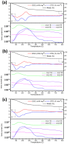
TG, DTG, and DSC curves (top) and gas products (bottom) of La/GNFs_ox (a), Nd/GNFs_ox (b), and Gd/GNFs_ox (c) heat treatments under Ar atmosphere.
Figure 5
Figure 5
X-ray data of GNFs, GNFs_ox, and Ln/CNFs_ox.
Figure 6
Figure 6
HRTEM (top row), HAADF-STEM (middle row) images and EELS spectra (lower row) of Ln/CNFs_ox particles: La/CNFs_ox (a,d), Nd/CNFs_ox (b,e), and Gd/CNFs_ox (c,f).
Figure 7
Figure 7
Survey XPS (a); La3d, Nd3d, Gd3d (b); O1s (c) and C1s (d) spectra of samples Ln/CNFs_ox.
Figure 8
Figure 8
(a) CT image of the phantom at 45 thl, (b) dependence of gamma radiation absorption on the energy (detector threshold) in the ROI, (c) application of the criterion, and (d) evaluation of the molar concentration of the CA. Water solutions with the following concentrations of the contrasting elements were used: iodine—140 mg·mL−1, Gd—74 mg·mL−1, La—40 mg·mL−1, and Nd—40 mg·mL−1.
Figure 9
Figure 9
Upper row: CT images of phantom with the standard samples (water, iodine, paraffin, bone, and plex) and the investigated water solutions of La(NO3)3·6H2O (a), Nd(NO3)3·6H2O (b), and Gd(NO3)3·6H2O (c). The numbers shown in the images reflect the concentration of the contrasting element in the study solution. Second row: results of application of the photon energy criteria. Green corresponds to La (d), Nd (e), and Gd (f), while yellow corresponds to samples with no contrasting elements detected by criteria. Third row: the estimation of molar concentrations of La (g), Nd (h), and Gd (i). Bottom row: dependence of simulated concentration criteria from experimental concentrations of water solutions of La(NO3)3·6H2O (j), Nd(NO3)3·6H2O (k), and Gd(NO3)3·6H2O (l).
Figure 10
Figure 10
The 3D CT images of phantom with Ln/GNFs_ox (a) and color division of La/GNFs_ox (red), Nd/GNFs_ox (blue), and Gd/GNFs_ox (green) according to criteria (b).

References

    1. Lusic H., Grinstaff M.W. X-Ray-Computed Tomography Contrast Agents. Chem. Rev. 2013;113:1641–1666. doi: 10.1021/cr200358s. - DOI - PMC - PubMed
    1. Wallyn J., Anton N., Akram S., Vandamme T.F. Biomedical Imaging: Principles, Technologies, Clinical Aspects, Contrast Agents, Limitations and Future Trends in Nanomedicines. Pharm. Res. 2019;36:78. doi: 10.1007/s11095-019-2608-5. - DOI - PubMed
    1. Hsu J.C., Nieves L.M., Betzer O., Sadan T., Noël P.B., Popovtzer R., Cormode D.P. Nanoparticle Contrast Agents for X-ray Imaging Applications. WIREs Nanomed. Nanobiotechnol. 2020;12:e1642. doi: 10.1002/wnan.1642. - DOI - PMC - PubMed
    1. Ostadhossein F., Tripathi I., Benig L., LoBato D., Moghiseh M., Lowe C., Raja A., Butler A., Panta R., Anjomrouz M., et al. Multi-“Color” Delineation of Bone Microdamages Using Ligand-Directed Sub-5 Nm Hafnia Nanodots and Photon Counting CT Imaging. Adv. Funct. Mater. 2020;30:1904936. doi: 10.1002/adfm.201904936. - DOI
    1. Anderson N.G., Butler A.P., Scott N.J.A., Cook N.J., Butzer J.S., Schleich N., Firsching M., Grasset R., de Ruiter N., Campbell M., et al. Spectroscopic (Multi-Energy) CT Distinguishes Iodine and Barium Contrast Material in MICE. Eur. Radiol. 2010;20:2126–2134. doi: 10.1007/s00330-010-1768-9. - DOI - PubMed

LinkOut - more resources