. 2022 Dec 5;11(23):3385.
doi: 10.3390/plants11233385.

DNA Barcoding and Morphometry Reveal Further Cryptic Bio-Diversity within the Pin Nematode Genus Paratylenchus (Nematoda: Tylenchulidae)

Affiliations

DNA Barcoding and Morphometry Reveal Further Cryptic Bio-Diversity within the Pin Nematode Genus Paratylenchus (Nematoda: Tylenchulidae)

Juan Emilio Palomares-Rius et al. Plants (Basel). .

Abstract

Paratylenchus species are obligate ectoparasitic nematodes on cultivated and wild herbaceous and woody plants occupying numerous soil categories. Several species may cause damage to several crops (viz. P. dianthus, P. enigmaticus, P. microdorus, P. hamatus and P. epacris on carnation, lettuce, rose and walnut, respectively). This investigation proves and emphasizes the relevance of applying integrative taxonomy for the accurate detection of Paratylenchus species in mountainous wild environments in the Malaga province, Southern Spain. This research analyzed 45 soil samples of maritimus pine and one of green heather in southern Spain and identified fourteen Paratylenchus species, two of them are described herein as new species (P. paraaonli sp. nov., P. plesiostraeleni sp. nov.), six of them were first reports for Spain (P. canchicus, P. nainianus, P. neonanus, P. salubris, Paratylenchus sp. 2 SAS, and P. wuae), and six species (P. caravaquenus, P. microdorus, P. nanus, P. neoamblycephalus, P. sheri, and P. variabilis) have been already reported in Spain. Accordingly, these data increase the biodiversity of pin nematodes in Spain comprising a total of 47 species (33.1% out of 142 total species of this genus). Phylogenetic analyses based on ribosomal and mitochondrial markers (D2-D3, ITS, and partial COI) resulted in a consistent position for the newly described Paratylenchus species in this study (P. plesiostraeleni sp. nov., P. paraaonli sp. nov.). Paratylenchus plesiostraeleni sp. nov. grouped in a separated subclade as unequivocal species from the P. straeleni-complex species (including P. straeleni and P. parastraeleni), and P. paraaonli sp. nov. clustered with P. vitecus, but clearly separate from this species. This study indicates that Paratylenchus species diversity in natural environments may be higher than expected, and this study may help in accurate identifications.

Keywords: D2-D3 expansion domains of the large ribosomal subunit (28S); cryptic species; cytochrome c oxidase c subunit 1 (COI); internal transcribed spacer (ITS); new species.


Conflict of interest statement

The authors declare no conflict of interest. The funders had no role in the design of the study; in the collection, analyses, or interpretation of data; in the writing of the manuscript, or in the decision to publish the results.

Figures
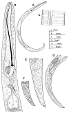
Figure 1
Figure 1
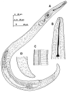
Line illustrations of Paratylenchus paraaonli sp. nov. (A) Whole female body; (B) Female pharyngeal region; (C) Detail of lateral field at mid-body; (D) Female posterior region; (E,F) Female tail.
Figure 2
Figure 2
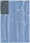
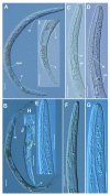
Micro-photomicrographs of Paratylenchus paraaonli sp. nov. female. (A) Whole female with stylet and vulva arrowed; (B) pharyngeal region; (C,D) detail of female stylet region; (E) detail of lateral fields; (FH) female posterior region with vulva and anus (arrowed) and detail of vulva showing advulval flap (arrowed); (I) female tail; (J) detail of spermatheca (arrowed). Scale bars (A = 50 µm; BJ = 20 µm). (Abbreviations: a = anus; avf = advulval flap; ep = excretory pore; lf = lateral field; st = stylet; spm = spermatheca; st = stylet; V = vulva).
Figure 3
Figure 3
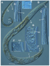
Micro-photomicrographs of Paratylenchus paraaonli sp. nov. third- and fourth-stage juveniles. (A,B) Entire third-stage juveniles showing stylet (arrowed), and insert of pharyngeal region and tail; (C) Entire fourth-stage juvenile showing pharyngeal region without stylet. Scale bars (AC = 20 µm). (Abbreviations: a = anus; st = stylet).
Figure 4
Figure 4
Line illustrations of Paratylenchus plesiostraeleni sp. nov. (A) Whole female; (B) Female stylet region; (C) Detail of lateral fields at mid-body; (D) Female tail.
Figure 5
Figure 5
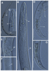
Micro-photomicrographs of Paratylenchus plesiostraeleni sp. nov. female. (A,B) Whole female with stylet, excretory pore and vulva arrowed; (C) pharyngeal region; (D) Stylet region; (E) Detail of lateral fields; (F) Detail of spermatheca; (G) Detail of vulva showing advulval flap (arrowed) and egg (arrowed). Scale bars (AG = 20 µm). (Abbreviations: a = anus; avf = advulval flap; egg = egg; ep = excretory pore; lf = lateral field; spm = spermatheca; st = stylet; V = vulva).
Figure 6
Figure 6
Micro-photomicrographs of Paratylenchus plesiostraeleni sp. nov. third- and fourth-stage juveniles. (A) Whole third-stage juvenile showing stylet (arrowed); (B) Pharyngeal region showing stylet (arrowed); (C) Whole fourth-stage juvenile showing absence of stylet and few developed pharynxes (arrowed). (D) Pharyngeal region of fourth-stage juvenile showing non-functional pharynx. Scale bars (AD = 20 µm). (Abbreviations: ph = pharynx; st = stylet).
Figure 7
Figure 7
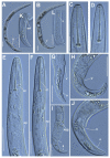
Micro-photomicrographs of Paratylenchus canchicus Mohilal and Dhanachand, 2004 female. (A) Whole female with vulva arrowed; (B,C) Pharyngeal region; (D) Lip region; (E) Female posterior region with vulva and anus (arrowed) and detail of vulva showing advulval flap membrane (arrowed). Scale bars (AC,E = 20 µm; D = 10 µm). (Abbreviations: a = anus; avf = advulval flap; dgo = dorsal gland orifice; st = stylet; V = vulva).
Figure 8
Figure 8
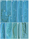
Micro-photomicrographs of Paratylenchus nanus Cobb, 1923 female. (A) Whole female with excretory pore and vulva arrowed; (B) Pharyngeal region with excretory pore arrowed; (C) Detail of stylet region; (D,E) Female posterior region with vulva and anus (arrowed) and detail of vulva showing advulval flap membrane (arrowed); (F) detail of lateral fields; (G) Detail of female tail. Scale bars (A,B,D,G = 20 µm; C,E,F = 10 µm). (Abbreviations: a = anus; avf = advulval flap; ep = excretory pore; lf = lateral field; st = stylet; V = vulva).
Figure 9
Figure 9
Micro-photomicrographs of Paratylenchus nainianus Edward & Misra, 1963 female. (A,B) Whole female with vulva arrowed; (C,D) Detail of female stylet region; (E,F) Pharyngeal region; (GK) Female posterior region with vulva and anus (arrowed) and detail of vulva showing advulval flap (arrowed); (L) Detail of lateral fields (lines arrowed). Scale bars (A,B,EL = 20 µm; C,D = 10 µm). (Abbreviations: a = anus; avf = advulval flap; egg = egg, ep = excretory pore; lf = lateral field; st = stylet; V = vulva).
Figure 10
Figure 10
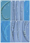
Micro-photomicrographs of Paratylenchus neonanus Mathur, Khan & Prasad, 1967 female and male. (A) Whole female with stylet and vulva arrowed; (B–E) Female pharyngeal region with stylet and excretory pore arrowed; (F) Female lip region; (G–I) Female posterior region with vulva and Postvulval uterine sac (arrowed) and detail of vulva showing advulval flap (arrowed); (J) Whole male; (K) Male pharyngeal region showing absence of stylet; (L) Male posterior region showing spicules (arrowed). Scale bars (A–L = 20 µm). (Abbreviations: avf = advulval flap; ep = excretory pore; lf = lateral field; pus = post-vulval uterine sac; sp = spicules; st = stylet; V = vulva).
Figure 11
Figure 11
Micro-photomicrographs of Paratylenchus salubris Raski, 1975 female. (A) Whole female with stylet, excretory pore and vulva arrowed; (B–D) Pharyngeal region; (E) Detail of lateral fields; (FI) Female posterior region with vulva and anus (arrowed) and detail of vulva showing advulval flap (arrowed). Scale bars (AI = 20 µm). (Abbreviations: a = anus; avf = advulval flap; ep = excretory pore; lf = lateral field; st = stylet; V = vulva).
Figure 12
Figure 12
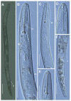
Micro-photomicrographs of Paratylenchus sp. 2 SAS female. (A) Whole female with vulva arrowed; (B,C) Pharyngeal region; (D,E) Detail of female stylet region; (F) Female posterior region with vulva (arrowed). Scale bars (A = 50 µm; B–F = 20 µm). (Abbreviations: dgo = dorsal gland orifice; ep = excretory pore; st = stylet; V = vulva).
Figure 13
Figure 13
Micro-photomicrographs of Paratylenchus wuae Yu, Ye & Powers, 2016 female. (A,B) Whole female with stylet, vulva and spermatheca arrowed; (C–G) Pharyngeal and stylet regions; (H) Detail of lateral fields; (IJ) Female posterior region with vulva and anus (arrowed). Scale bars (AG,I,J = 20 µm; H = 10 µm). (Abbreviations: a = anus; ep = excretory pore; lf = lateral field; spm = spermatheca; st = stylet; V = vulva).
Figure 14
Figure 14
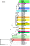
Phylogenetic relationships within the genus Paratylenchus. Bayesian 50% majority rule consensus tree as completed from D2-D3 expansion domains of the 28S rRNA sequence alignment under the general time-reversible model with invariable sites and gamma distribution model (GTR + I + G). Posterior probabilities of more than 0.70 are given for appropriate clades. Newly obtained sequences in this study are shown in bold. The scale bar indicates expected changes per site, and the colored boxes indicate the clade association of new Paratylenchus species sequenced in this study.
Figure 15
Figure 15
Phylogenetic relationships within the genus Paratylenchus. Bayesian 50% majority rule consensus tree as completed from ITS rRNA sequence alignment under the general time-reversible model with invariable sites and gamma distribution model (GTR + I + G). Posterior probabilities of more than 0.70 are given for appropriate clades. Newly obtained sequences in this study are shown in bold. The scale bar indicates expected changes per site, and the colored boxes indicate the clade association of new Paratylenchus species sequenced in this study.
Figure 16
Figure 16
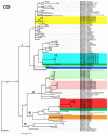
Phylogenetic relationships within the genus Paratylenchus. Bayesian 50% majority rule consensus tree as completed from cytochrome c oxidase subunit 1 (COI) sequence alignment under the general time-reversible model with invariable sites and gamma distribution model (GTR + I + G). Posterior probabilities of more than 0.70 are given for appropriate clades. Newly obtained sequences in this study are shown in bold. The scale bar indicates expected changes per site, and the colored boxes indicate the clade association of new Paratylenchus species sequenced in this study.

References

    1. Micoletzky H. Die freilebenden Erd-Nematoden. Arch. Für Nat. 1922;87:1–650.
    1. Ghaderi R., Geraert E., Karegar A. The Tylenchulidae of the World, Identification of the Family Tylenchulidae (Nematoda: Tylenchida) Academia Press; Ghent, Belgium: 2016.
    1. Singh R., Karssen G., Coureur M., Subbotin S., Bert W. Integrative taxonomy and molecular phylogeny of the plant-parasitic nematode genus Paratylenchus (Nematoda: Paratylenchinae): Linking species with molecular barcodes. Plants. 2021;10:408. doi: 10.3390/plants10020408. - DOI - PMC - PubMed
    1. Clavero-Camacho I., Cantalapiedra-Navarrete C., Archidona-Yuste A., Castillo P., Palomares-Rius J.E. Remarkable cryptic diversity of Paratylenchus spp. (Nematoda: Tylenchulidae) in Spain. Animals. 2021;11:1161. doi: 10.3390/ani11041161. - DOI - PMC - PubMed
    1. Clavero-Camacho I., Palomares-Rius J.E., Cantalapiedra-Navarrete C., Leon-Ropero G., Martin-Barbarroja J., Archidona-Yuste A., Castillo P. Integrative taxonomy reveals hidden cryptic diversity within pin nematodes of the genus Paratylenchus (Nematoda: Tylenchulidae) Plants. 2021;10:1454. doi: 10.3390/plants10071454. - DOI - PMC - PubMed

LinkOut - more resources