. 2023 Apr;20(2):213-223.
doi: 10.1007/s13770-022-00504-x. Epub 2022 Dec 11.

Tissue Engineered Mini-Cornea Model for Eye Irritation Test

Affiliations

Tissue Engineered Mini-Cornea Model for Eye Irritation Test

Seon-Hwa Kim et al. Tissue Eng Regen Med. 2023 Apr.

Abstract

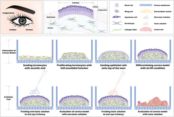
Background: Eye irritation tests with animals have been conducted for a long time. However, the subjective decision to irritation, the anatomic/physiologic difference between species and humans, and ethical issues are crucial problems. Various research groups have paid attention to alternative testing methods. In these senses, we fabricated in vitro mini-cornea models with immortalized human corneal epithelial cells (iHCECs) and keratocytes (iHCKs) and used them for irritation tests. This study hypothesized that our mini-cornea model could present different viability tendencies according to test chemicals with different irritancy levels.

Methods: Cells used in this study were characterized with cornea-specific markers by immunocytochemistry and western blot. To make a three-dimensional hemisphere construct like cornea stroma, we cultured iHCKs under modified culture conditions verified by matrix formation and total collagen content. iHCECs were seeded on the construct and cultured at an air-liquid interface. The model was treated with 2-phenoxyethanol, triton X-100, sodium lauryl sulfate, and benzalkonium chloride.

Results: iHCECs and iHCKs presented their specific cell markers. In modifying the culture condition, the group treating ascorbic acid (200 µg/ml) presented an intact cellular matrix and included the highest collagen content; thus, we used this condition to fabricate the mini-cornea model. The model shows hemisphere shape and homogenous cell distributions in histological analysis. We observed different sensitivity tendencies by types of chemicals, and the model's viability significantly decreased when the chemical concentration increased.

Conclusion: In this study, we performed and observed irritation tests using a tissue-engineered mini-cornea model and considered to apply as an alternative approach for animal tests.

Keywords: Ascorbic acid; Eye irritation; In vitro cornea model; Tissue engineering.


Conflict of interest statement

The authors declare they have no conflicts of interest.

Figures

Fig. 1
Fig. 1
Schematic illustration of fabricating and evaluating the mini-cornea model
Fig. 2
Fig. 2
Cornea-related protein marker expression in immortalized human corneal epithelial cells (iHCECs) and keratocytes (iHCKs). A Western blotting analysis of iHCECs and iHCKs combined with CK 18, IL-4Rα, IL-17R, and CK 19. GAPDH was used as a housekeeping protein. B Immunofluorescent images of iHCECs and iHCKs stained with cornea-related markers (red), F-actin (green), and nuclei (blue) (Scale bar: 20 µm)
Fig. 3
Fig. 3
Formation of synthetic matrices as 2D sheets by iHCKs and quantitating total collagen content of the sheets. All groups were presented to control (0 µg/ml of ascorbic acid), low (50 µg/ml), mid (100 µg/ml), and high (200 µg/ml). A Microscopic images were captured when the sheets were under conditions that attached (left) to the surfaces and detached (right) from the culture vessels. All sheets were detached with micropipette after culture 48 h. The white solid and dotted line show each edge and folded margin of fabricated sheets (Scale bar: 200 µm). Macroscopic images indicate the whole appearance of the sheets, and magnified images in specific regions were marked with square boxes (red). The S and D indicate the region of the cell sheet and culture dish. B Graph for total collagen content of synthesized cell matrix cultured for 6 days. Data were presented as mean ± SEM (n = 3/group) with statistical significance (*p < 0.05 and ***p < 0.001)
Fig. 4
Fig. 4
Histology and immunostaining to compare corneal tissue and mini-cornea model. A H&E staining images of rat cornea (top) and the mini-cornea model (bottom). B Immunohistochemistry images of rat cornea (top) and the mini-cornea model (bottom) with CK 3, CK 18, E-Cad, and VI (Scale bar: 100 µm). The blow-up images in boundary regions consisting of iHCECs and iHCKs were indicated with the square box (red)
Fig. 5
Fig. 5
Evaluating cell viability in the mini-cornea model for eye irritation test A 2-Phenoxyethanol (PHE)-treated group with concentrations of 0, 0.3, 0.6, 1, and 2%. B Triton X-100 (TX-100)-treated group with concentrations of 0, 0.1, 0.3, 0.45, and 1%. C Sodium lauryl sulfate (SLS)-treated group with concentrations of 0, 0.1, 0.3, 0.6, and 1%. D Benzalkonium chloride (BAK)-treated group with concentrations of 0, 0.01, 0.03, 0.045, and 0.06%. Measured values were presented to mean ± SEM (n = 4/group) and statistical significance (*p < 0.05, **p < 0.01, and ***p < 0.001)
Fig. 6
Fig. 6
Histological observation of the mini-cornea model treated with HBSS, 2% PHE, 0.6% TX-100, 1% SLS, and 0.06% BAK. Magnifying images show the morphologies of each testing solution-exposed area (Scale bar: 100 µm)

Similar articles

Cited by

References

    1. Abou Shousha M, Wang J, Kontadakis G, Feuer W, Canto AP, Hoffmann R, et al. Corneal epithelial thickness profile in dry-eye disease. Eye (Lond) 2020;34:915–922. doi: 10.1038/s41433-019-0592-y. - DOI - PMC - PubMed
    1. DelMonte DW, Kim T. Anatomy and physiology of the cornea. J Cataract Refract Surg. 2011;37:588–598. doi: 10.1016/j.jcrs.2010.12.037. - DOI - PubMed
    1. Lagali N. Corneal stromal regeneration: current status and future therapeutic potential. Curr Eye Res. 2020;45:278–290. doi: 10.1080/02713683.2019.1663874. - DOI - PubMed
    1. Syed-Picard FN, Du Y, Hertsenberg AJ, Palchesko R, Funderburgh ML, Feinberg AW, et al. Scaffold-free tissue engineering of functional corneal stromal tissue. J Tissue Eng Regen Med 2018; 12:59–69. - PMC - PubMed
    1. Sridhar MS. Anatomy of cornea and ocular surface. Indian J Ophthalmol. 2018;66:190–194. doi: 10.4103/ijo.IJO_646_17. - DOI - PMC - PubMed

Publication types

LinkOut - more resources