Review
. 2022 Jan-Dec;14(1):2154548.
doi: 10.1080/19490976.2022.2154548.

From intestinal colonization to systemic infections: Candida albicans translocation and dissemination

Affiliations
Review

From intestinal colonization to systemic infections: Candida albicans translocation and dissemination

Jakob L Sprague et al. Gut Microbes. 2022 Jan-Dec.

Abstract

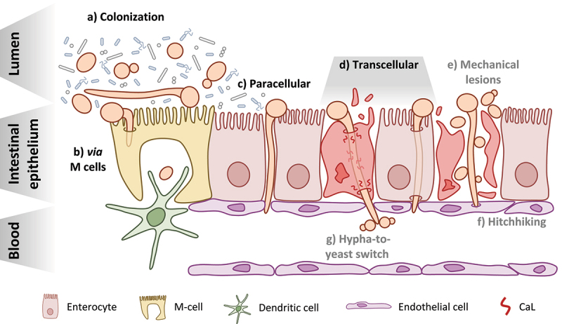
Candida species are the most prevalent cause of invasive fungal infections, of which Candida albicans is the most common. Translocation across the epithelial barrier into the bloodstream by intestinal-colonizing C. albicans cells serves as the main source for systemic infections. Understanding the fungal mechanisms behind this process will give valuable insights on how to prevent such infections and keep C. albicans in the commensal state in patients with predisposing conditions. This review will focus on recent developments in characterizing fungal translocation mechanisms, compare what we know about enteric bacterial pathogens with C. albicans, and discuss the different proposed hypotheses for how C. albicans enters and disseminates through the bloodstream immediately following translocation.

Keywords: Candida albicans; dissemination; fungi; gut colonization; intestinal translocation.


Conflict of interest statement

No potential conflict of interest was reported by the authors.

Figures

Figure 1.
Figure 1.
Candida albicans at the intestinal barrier: from colonization to translocation and dissemination. a) C. albicans can be found in both the yeast and hyphal morphotypes during colonization of the GI tract of healthy individuals and must compete with other members of the microbiota b) Translocation via M cells present in intestinal lymphoid tissues like Peyer’s patches may occur following either induced endocytosis or active penetration by C. albicans and may also involve the fungus hijacking the sampling function of resident phagocytes, like dendritic cells c) The paracellular route of translocation refers to invasion of the intestinal barrier by C. albicans hyphae in the spaces between enterocytes without actually invading the host cells d) The transcellular route of translocation could occur in two manners: with candidalysin (CaL)-dependent damage of the epithelial cells resulting in necrotic cell death (left) or in the absence of host-cell damage (right) This appears to be the major route used by C. albicans to cross the intestinal barrier e) One hypothesis for how yeast reach the bloodstream is that they are able to move across the epithelial barrier from the GI tract to the bloodstream via physical disruptions in the tissue, caused either by the fungus itself or some other factors like surgery or chemotherapy f) The so-called “hitchhiking” hypothesis posits that yeast present in the GI tract may move through the barrier along with invading hyphae by attaching to them as they invade the epithelium g) Finally, the hypha-to-yeast switch hypothesizes that translocating hyphae undergo a morphological transition once reaching the bloodstream that initiates the formation of yeast cells from the hyphae.
Figure 2.
Figure 2.
A glance at intestinal epithelial cell junctions. An overview of the major components of cell-cell junctions of enterocytes Tight junctions (TJ) are mainly comprised of the occludin, claudin, and junction adhesion molecule (JAM) proteins Adherens junctions (AJ) in intestinal epithelial cells are mostly comprised of E-cadherin, catenins, and nectins C. albicans is capable of degrading tight and adherens junction proteins, potentially via secretion of Sap5.

References

    1. Kullberg BJ, Arendrup MC, Campion EW.. Invasive Candidiasis. N Engl J Med. Oct 8 2015;373(15):1445–14. doi: 10.1056/NEJMra1315399 - DOI - PubMed
    1. Pappas PG, Lionakis MS, Arendrup MC, Ostrosky-Zeichner L, Kullberg BJ. Invasive candidiasis. Nat Rev Dis Primers May. 2018;11(4):18026. doi: 10.1038/nrdp.2018.26. - DOI - PubMed
    1. Ruhnke M, Groll AH, Mayser P, Ullmann AJ, Mendling W, Hof H, Denning DW. 2015. Denning DW estimated burden of fungal infections in Germany. Mycoses. Oct;58(Suppl 5):22–28. doi: 10.1111/myc.12392. - DOI - PubMed
    1. Nash AK, Auchtung TA, Wong MC, Smith DP, Gesell JR, Ross MC, Stewart CJ, Metcalf GA, Muzny DM, Gibbs RA, et al. The gut mycobiome of the Human Microbiome Project healthy cohort. Microbiome. Nov 25 2017;5(1):153. 10.1186/s40168-017-0373-4. PMC5702186. - DOI - PMC - PubMed
    1. Reagan DR, Pfaller MA, Hollis RJ, Wenzel RP. Wenzel RP Characterization of the sequence of colonization and nosocomial candidemia using DNA fingerprinting and a DNA probe. J Clin Microbiol Dec. 1990;28(12):2733–2738. doi: 10.1128/jcm.28.12.2733-2738.1990. PMC268264 - DOI - PMC - PubMed

LinkOut - more resources