

Comparison of adhesion of thawed and cultured synovial mesenchymal stem cells to the porcine meniscus and the relevance of cell surface microspikes
- PMID: 36503422
- PMCID: PMC9743635
- DOI: 10.1186/s12860-022-00456-z
Comparison of adhesion of thawed and cultured synovial mesenchymal stem cells to the porcine meniscus and the relevance of cell surface microspikes
Abstract
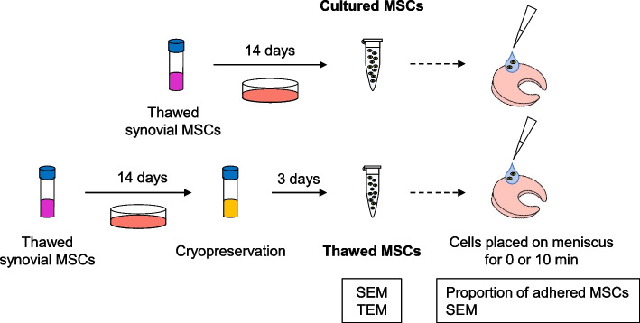
Background: Placement of a cultured synovial mesenchymal stem cell (MSC) suspension on a repaired meniscus for 10 min accelerated meniscus repair. Upon placement of the MSC suspension on the meniscus, microspikes projecting from the MSC surface trap meniscus fibers and promote MSC adhesion. Thawed cryopreserved MSCs are preferred materials for meniscus repair, as they can be transplanted without additional culture. However, the adhesion ability of thawed cryopreserved MSCs is unknown. Here, we compared the proportion of cultured versus thawed MSCs adhering to a porcine meniscus immediately and 10 min after placement. We also investigated the relationship between adhesion and the number of microspikes on the synovial MSCs.
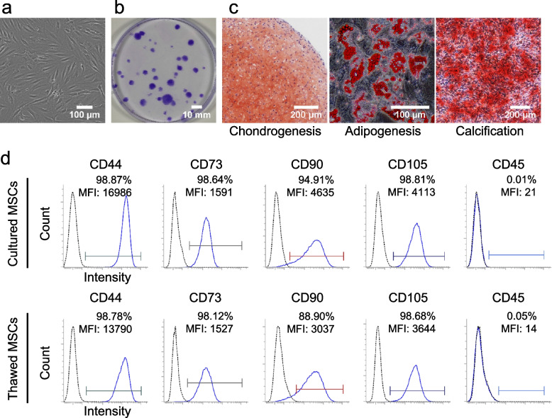
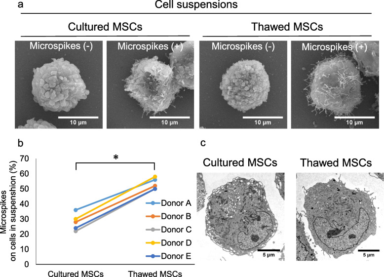
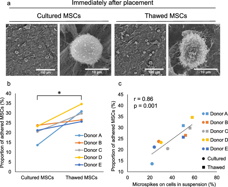
Methods: Synovial MSCs were prepared from the knees of four donors with osteoarthritis. The "cultured MSCs" were thawed MSCs that were re-cultured and suspended in PBS for transplantation. A similarly prepared suspension was cryopreserved, thawed again, suspended in PBS, and used without further culture as the "thawed MSCs." MSCs with at least three microspikes in SEM images were defined as microspike-positive MSCs. Porcine meniscus surfaces were abraded, cut into a cylindrical shape, and treated with MSC suspension. Non-adherent cells were counted immediately and again 10 min after placement to calculate the adhesion proportion.
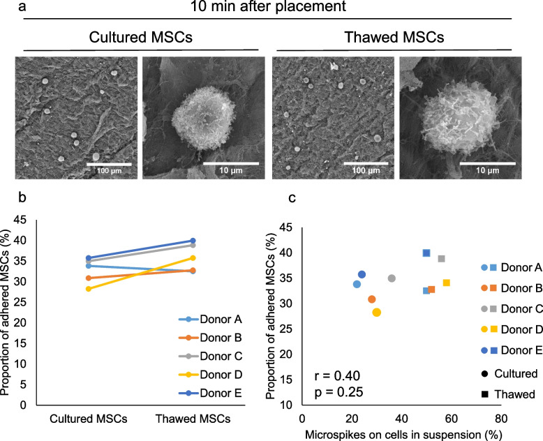
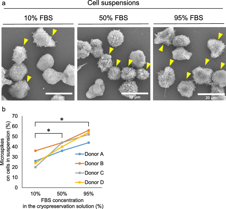
Results: The proportion of microspike-positive MSCs was significantly higher in thawed (53 ± 3%) than in cultured (28 ± 5%) MSC suspensions. MSC adhesion to the meniscus was significantly better for the thawed than for the cultured MSC suspensions immediately after placement on the meniscus, but no differences were detected after 10 min. The proportion of MSCs with microspikes in the cell suspension was significantly correlated with the proportion of adhered MSCs immediately after the placement, but not 10 min later. Addition of FBS to the cryopreservation medium promoted a concentration-dependent increase in the proportion of microspike-positive cells.
Conclusions: Thawed MSCs adhered better than cultured MSCs immediately after placement, but adhesion was similar for both MSC preparations after 10 min. Immediately after placement, the presence of microspikes was associated with better adhesion of synovial MSCs to the meniscus.
Keywords: Adhesion; Cryopreservation; Meniscus; Mesenchymal stem cell; Microspike; Scanning electron microscopy; Synovium; Thawed cell.
© 2022. The Author(s).
Conflict of interest statement
The authors declare that they have no conflicts of interest.
Figures
Similar articles
-
Scanning electron microscopy analysis of synovial and adipose mesenchymal stem cells adhering to cartilage.Regen Ther. 2024 May 5;27:488-495. doi: 10.1016/j.reth.2024.04.012. eCollection 2024 Dec. Regen Ther. 2024. PMID: 38756702 Free PMC article.
-
Morphological changes in synovial mesenchymal stem cells during their adhesion to the meniscus.Lab Invest. 2020 Jul;100(7):916-927. doi: 10.1038/s41374-020-0421-8. Epub 2020 Apr 1. Lab Invest. 2020. PMID: 32238905
-
Human synovial mesenchymal stem cells show time-dependent morphological changes and increased adhesion to degenerated porcine cartilage.Sci Rep. 2022 Oct 5;12(1):16619. doi: 10.1038/s41598-022-20386-2. Sci Rep. 2022. PMID: 36198727 Free PMC article.
-
[Homeostasis and Disorder of Musculoskeletal System.Transplantation of synovial mesenchymal stem cells for cartilage and meniscus regeneration.].Clin Calcium. 2018;28(3):319-327. Clin Calcium. 2018. PMID: 29512522 Review. Japanese.
-
Synovial membrane mesenchymal stem cells for cartilaginous tissues repair.Mol Biol Rep. 2022 Mar;49(3):2503-2517. doi: 10.1007/s11033-021-07051-z. Epub 2022 Jan 11. Mol Biol Rep. 2022. PMID: 35013859 Review.
Cited by
-
Scanning electron microscopy analysis of synovial and adipose mesenchymal stem cells adhering to cartilage.Regen Ther. 2024 May 5;27:488-495. doi: 10.1016/j.reth.2024.04.012. eCollection 2024 Dec. Regen Ther. 2024. PMID: 38756702 Free PMC article.
References
MeSH terms
Grants and funding
LinkOut - more resources
Full Text Sources
