. 2022 Dec 12;18(1):134.
doi: 10.1186/s13007-022-00963-2.

A low-cost and open-source solution to automate imaging and analysis of cyst nematode infection assays for Arabidopsis thaliana

Affiliations

A low-cost and open-source solution to automate imaging and analysis of cyst nematode infection assays for Arabidopsis thaliana

Olaf Prosper Kranse et al. Plant Methods. .

Abstract

Background: Cyst nematodes are one of the major groups of plant-parasitic nematode, responsible for considerable crop losses worldwide. Improving genetic resources, and therefore resistant cultivars, is an ongoing focus of many pest management strategies. One of the major bottlenecks in identifying the plant genes that impact the infection, and thus the yield, is phenotyping. The current available screening method is slow, has unidimensional quantification of infection limiting the range of scorable parameters, and does not account for phenotypic variation of the host. The ever-evolving field of computer vision may be the solution for both the above-mentioned issues. To utilise these tools, a specialised imaging platform is required to take consistent images of nematode infection in quick succession.

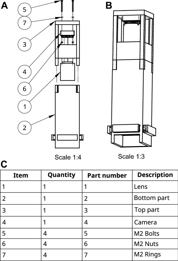
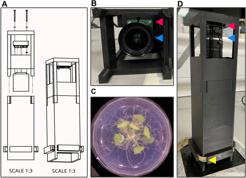
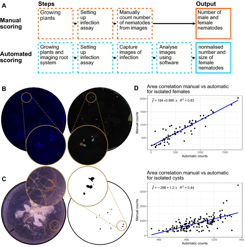
Results: Here, we describe an open-source, easy to adopt, imaging hardware and trait analysis software method based on a pre-existing nematode infection screening method in axenic culture. A cost-effective, easy-to-build and -use, 3D-printed imaging device was developed to acquire images of the root system of Arabidopsis thaliana infected with the cyst nematode Heterodera schachtii, replacing costly microscopy equipment. Coupling the output of this device to simple analysis scripts allowed the measurement of some key traits such as nematode number and size from collected images, in a semi-automated manner. Additionally, we used this combined solution to quantify an additional trait, root area before infection, and showed both the confounding relationship of this trait on nematode infection and a method to account for it.

Conclusion: Taken together, this manuscript provides a low-cost and open-source method for nematode phenotyping that includes the biologically relevant nematode size as a scorable parameter, and a method to account for phenotypic variation of the host. Together these tools highlight great potential in aiding our understanding of nematode parasitism.


Conflict of interest statement

The authors are not aware of competing interests.

Figures

Fig. 1
Fig. 1
Explosion diagram of the imaging tower. A The separate components labelled with their corresponding end position. The item numbers indicate a position in the table below. B The final product after assembly. C A table of components. The item number corresponds with the numbers in A
Fig. 2
Fig. 2
An example of a manual outline drawn around a female cyst nematode using ImageJ. (Left) before drawing an outline. (Right) the outline drawn
Fig. 3
Fig. 3
The imaging tower. A An explosion diagram of the assembly and final product of the imaging tower on scale of 1:3. B a look inside the top half of the imaging tower. C An example image taken using the imaging tower. D The assembled imaging tower. The lens (blue arrow) is mounted to the camera (red arrow), which is connected to the Raspberry Pi (not shown). Consistent all around lighting is provided by an LED strip (yellow arrow)
Fig. 4
Fig. 4
Extraction of root surface area from images and correlation with nematode counts. A A visual representation of the pipeline used for calculating the root surface area. From left to right the features are extracted from the image and a region of interest is defined. Artefacts picked up in the pipeline: A1 Damage to the agar (purple arrow) and a leaf touching the agar (brown arrow). A2 A small particle on the surface of the plastic (pink arrow), clustered roots interpreted as a singular root (blue arrow). B An example of the ability to extract the leaf surface area using colour thresholding. Artifacts are introduced by difference in leaf colour (pink arrow) and damage to the agar (blue arrow). C Two models describing the relationship between number of nematodes and root surface area for two A. thaliana genotypes. The models are described as follows; Columbia 0, Males: y = 5.4 + 5.7 × 10−6x, Females: y = 17.5 + 6.33 × 10−6x. N804585, Males: y = 10 + 2.34 × 10−5x, Females: y = 11 + 1.26 × 10−5x
Fig. 5
Fig. 5
The spectrum of coloured agar under various lighting conditions. A The range of colours tested for contrast with nematodes and filtration potential of the background. B The colour red agar under four different lighting conditions. From left to right: White, Red, Green and Blue light. The blue lighting condition with the red agar were the most ideal combination for quantifying nematodes using the segmentation tool. The bottom image row has the contrast and brightness adjusted for visibility. Examples of female nematodes are highlighted with yellow arrows
Fig. 6
Fig. 6
Isolation and quantification of nematodes from images. A A schematic workflow highlighting the steps required for manual and automated nematode counting, and the differences in output. B Isolation of female cyst nematodes from red coloured agar under blue light illumination, and the extracted overlay from the script. C Isolation of encysted nematodes under white light illumination, and the extracted overlay from the script. D Correlation of area of the nematode between manual counts and automatic counts using the script for females and cysts

Similar articles

Cited by

References

    1. Abad P, Gouzy J, Aury JM, Castagnone-Sereno P, Danchin EGJ, Deleury E, et al. Genome sequence of the metazoan plant-parasitic nematode Meloidogyne incognita. Nat Biotechnol. 2008;26:909–915. doi: 10.1038/nbt.1482. - DOI - PubMed
    1. Sasser JN. A world perspective on nematology: the role of the society. Vistas Nematol. 1987;7–14.
    1. Chitwood DJ. Research on plant-parasitic nematode biology conducted by the United States Department of Agriculture-Agricultural Research Service. Pest Manag Sci. 2003;59:748–753. doi: 10.1002/ps.684. - DOI - PubMed
    1. Savary S, Willocquet L, Pethybridge SJ, Esker P, McRoberts N, Nelson A. The global burden of pathogens and pests on major food crops. Nat Ecol Evol. 2019;3:430–439. doi: 10.1038/s41559-018-0793-y. - DOI - PubMed
    1. Atkinson HJ, Urwin PE, Hansen E, McPherson MJ. Designs for engineered resistance to root-parasitic nematodes. Trends Biotechnol. 1995;13:369–374. doi: 10.1016/S0167-7799(00)88983-0. - DOI

LinkOut - more resources