Review
. 2023 May:94:101119.
doi: 10.1016/j.preteyeres.2022.101119. Epub 2022 Dec 8.

Circadian clock organization in the retina: From clock components to rod and cone pathways and visual function

Affiliations
Review

Circadian clock organization in the retina: From clock components to rod and cone pathways and visual function

Jacob D Bhoi et al. Prog Retin Eye Res. 2023 May.

Abstract

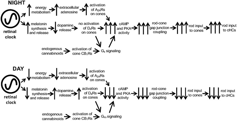
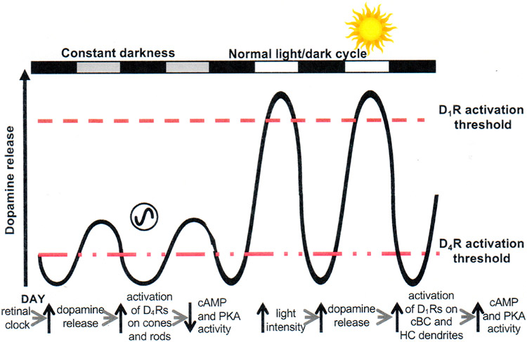
Circadian (24-h) clocks are cell-autonomous biological oscillators that orchestrate many aspects of our physiology on a daily basis. Numerous circadian rhythms in mammalian and non-mammalian retinas have been observed and the presence of an endogenous circadian clock has been demonstrated. However, how the clock and associated rhythms assemble into pathways that support and control retina function remains largely unknown. Our goal here is to review the current status of our knowledge and evaluate recent advances. We describe many previously-observed retinal rhythms, including circadian rhythms of morphology, biochemistry, physiology, and gene expression. We evaluate evidence concerning the location and molecular machinery of the retinal circadian clock, as well as consider findings that suggest the presence of multiple clocks. Our primary focus though is to describe in depth circadian rhythms in the light responses of retinal neurons with an emphasis on clock control of rod and cone pathways. We examine evidence that specific biochemical mechanisms produce these daily light response changes. We also discuss evidence for the presence of multiple circadian retinal pathways involving rhythms in neurotransmitter activity, transmitter receptors, metabolism, and pH. We focus on distinct actions of two dopamine receptor systems in the outer retina, a dopamine D4 receptor system that mediates circadian control of rod/cone gap junction coupling and a dopamine D1 receptor system that mediates non-circadian, light/dark adaptive regulation of gap junction coupling between horizontal cells. Finally, we evaluate the role of circadian rhythmicity in retinal degeneration and suggest future directions for the field of retinal circadian biology.

Keywords: Adenosine; Circadian clock; Circadian rhythmicity; Dopamine; Electrical synapses; Energy metabolism; Gap junctions; Melatonin; Retina; pH.


Figures

Fig. 1.
Fig. 1.. Schematic representation of a simplified circadian clock pathway.
(A) A simplified circadian clock pathway has three components. First, there is a molecular mechanism, which is referred to as a clock, clockwork, or a pacemaker, that has a period of approximately 24 hours and is able to maintain its daily rhythmicity in the absence of environmental cues (e.g., in constant darkness and temperature). Second, although circadian clocks can maintain rhythmicity in a constant environment, they are entrained or synchronized to the local environment by zeitgebers (German for time-giver). For example, the daily light/dark cycle acts as an input to clocks because the onset of morning light resets the “hands” of clocks so that they are set to local time, even though environments do not alter the period of clocks. Third, circadian clocks produce daily rhythms at the molecular, cellular, and systems levels. For example, the circadian clock in the retina produces a variety of daily rhythms within the retina, including increasing the hormone melatonin at night, decreasing the neurotransmitter dopamine in the day, and enabling cone photoreceptors to respond to very dim (scotopic) light stimuli at night (but not in the day). Because circadian clocks are affected by environmental stimuli, circadian experiments, which aim to determine whether time of day or night affects a function (e.g., level of extracellular dopamine), are conducted in the absence of environmental cues (e.g., under conditions of constant darkness and temperature). In a circadian experiment, the terms “subjective day” and “subjective night” refer to the day and night of the imposed light/dark cycle, respectively, when animals or isolated intact retinas were maintained in constant darkness. (B) Simplified representation of the core components of the mammalian circadian clockwork. See text for details.
Fig. 2.
Fig. 2.. A circadian clock regulates the light responses of fish cone horizontal cells (cHCs).
(A) Cone input to L-type cHCs predominates during the subjective day and rod input predominates during the subjective night. Compared to the day, the responses at night are slower, smaller in size, longer in duration and the response threshold is approximately a hundred times lower. Retinas were dark adapted for at least 1 h after excision, following which L-type cHCs were impaled without the aid of any light flashes. Responses of the cells to dim full-field white light flashes (ranging from −8 log Io to −5 log Io) were then recorded. The responses of two different cells are shown in the subjective day and night. (B) Average responses to a bright light stimulus (−3 log Io) as a function of time. Responses in the dark are greater during the subjective day than during the subjective night (open circles), even when the animals have been previously entrained to a reversed light/dark cycle (filled circles). The presence of a recording from a cHC was confirmed following cell impalement by flashing a series of dim (≤ −6 log Io) lights. Following this, a single bright (−3 log Io) light was flashed. Data were averaged only from responses to this single bright light stimulus (one bright light stimulus per retina). In each case, a response to a single bright light stimulus (−3 log Io) was obtained at the time indicated. (C) Spectral sensitivity measurements demonstrate a circadian rhythm of rod and cone input to cHCs. Average L-type cHC spectral sensitivity during the subjective night (~ CT15) resembles that of goldfish rod horizontal cells (and rods) (Schwanzara, 1967), rather than red (625 nm) cones (Harosi and MacNichol, 1974), for wavelengths ≤ 600 nm. In contrast, average L-type cHC spectral sensitivity during the subjective day (~ CT03) was similar to that of red (625 nm) cones. The relative spectral sensitivity of rod horizontal cells closely resembled that of goldfish rods, regardless of time of day (Ribelayga et al., 2007). Narrow band interference filters were used to control stimulus wavelength. Relative quantum sensitivity was determined using a 1 mV criterion response in order to minimize light sensitization of the dark-adapted state. Surgical isolation of the retina occurred approximately 2 h before this bright light response was recorded. Each data point represents averages obtained from 5-8 cells (1 cell per retina). Intensity values are relative to the maximum, unattenuated intensity (Io, 2.0 mW.cm−2) of full-field white light stimuli generated by the photostimulator. The maximum, unattenuated light intensity of the stimulus at 550 nm was 7.2 x 10¹³ photons.cm−2.sec−1. Adapted from Wang, Y., Mangel, S.C., 1996. Proc. Natl. Acad. Sci. USA 14, 4655-4660.
Fig. 3.
Fig. 3.. Circadian Variations in Cone Spectral Sensitivity, Light Response Threshold, and Receptive Field Size
(A) Average spectral sensitivity of cones recorded under dark-adapted conditions during the day or subjective day fit one of three nomograms (thin dotted curves) corresponding to the three major known types of goldfish cone pigments: L, M, and S. Data were obtained from recorded red cones (open squares; n = 9), green cones (open circles; n = 6) and blue cone (open triangle; n = 1). In contrast, the spectral sensitivity of all dark-adapted cones recorded at night peaked at ~ 535 nm (filled circles; n = 10). Although cone spectral sensitivity at night under dark-adapted conditions closely fits a rod nomogram (solid thick line) for 400 nm < λ < 600 nm, it does not fit the nomogram as well for λ > 600 nm. Rather, the data points closely fit a modified nomogram that combines goldfish rod and L-cone pigment nomograms (dotted thick curve; λmax = 537 ± 3 (s.d.) nm; r2 = 0.91). Following application of spiperone (10 μM) (open diamonds; n = 2), cone spectral sensitivity in the subjective day resembled that observed during the subjective night and data points fit well the modified nomogram (λmax = 537 ± 3 nm; r2 = 0.96). (B) Following bright light adaptation at night or during the subjective night 3 groups of cones with different spectral sensitivities were observed: red cones (filled squares; n = 4), green cones (filled circles; n = 5) and blue cone (filled triangles; n = 1), whereas bright light adaptation during the day or subjective day did not affect the relative spectral sensitivity of the recorded cones (red cones: open squares; n = 2; green cones: open circles; n = 6) but slightly decreased the absolute sensitivity. Nomograms as in (A). (A and B) Data points represent average sensitivity ± s.e.m. (C) Average day/night and circadian rhythms of the cone light response threshold (i.e. intensity required to elicit a 0.5 mV response) under dark-adapted conditions. The average cone light response threshold (log intensity) was significantly higher during the day (p < 0.001) and subjective day (p < 0.001) than during the night and subjective night (Tukey post hoc analysis). Data points represent averages of 4 to 15 measurements. (D) Average normalized response amplitudes of dark-adapted cones plotted against stimulus radius for a stimulus of intensity −5 log Io. These data indicate that the receptive field size of cones is larger at night than in the day. Measurements were performed during the day (open circles, n = 6) and night (filled circles, n = 6). (C, D) Error bars indicate s.e.m. Adapted from Ribelayga, C., Cao, Y., Mangel, S.C., 2008. Neuron 59, 790-801.
Fig. 4.
Fig. 4.. A circadian clock in the goldfish retina controls rod/cone gap junction coupling.
(A-D) Following iontophoresis of biocytin into individual cones, the tracer remained in a few cells (indicated by arrows in A1, D1) near the injected cone during the subjective day (A) and during the subjective night in the presence of the D4 receptor agonist quinpirole (1 μM, D), but diffused into many rods and cones during the subjective night (B) and during the subjective day in the presence of the D4 receptor antagonist spiperone (10 μM, C). In each of A-D, confocal images of a whole-mount retina at the level of the rod inner segments are shown on the left and perpendicular views of the 3-D reconstruction of the photoreceptor cells from the same retina are shown on the right. Some cones (arrows) and rods (arrowheads) are indicated. Scale bars (A-D): 50 μm. (E and F) Average numbers of stained cones (open bars) and rods (filled bars) following biocytin injections into individual cones (1 cone injected/retina) under dark-adapted conditions (E) during the day (n = 11) and subjective day (n = 5), night (n = 4) and subjective night (n = 5), subjective day in the presence of spiperone (n = 6), and subjective night in the presence of quinpirole (n = 6), and under dim light-adapted conditions (F-left) during the day (n = 6) and night (n = 3) and bright light-adapted conditions (F-right) during the day (n = 2) and night (n = 3). Under dark-adapted conditions, the number of tracer coupled rods and cones was significantly greater during the night (p < 0.001) and during the day following spiperone treatment (p < 0.001) than during the day under control conditions. Under dim light-adapted conditions, the number of tracer coupled rods and cones was significantly greater during the night (p < 0.001) compared to the day (Tukey post hoc analysis). Under bright light-adapted conditions, biocytin was restricted to the injected cone; no other cells were labeled. Error bars represent s.e.m. Adapted from Ribelayga, C., Cao, Y., Mangel, S.C., 2008. Neuron 59, 790-801.
Fig. 5.
Fig. 5.. Activation of dopamine D4, adenosine A2A, and cannabinoid CB1 receptors on rods and cones in the day and night work together to increase the day/night difference in rod/cone gap junction coupling.
The contribution of each receptor type (D4, A2A, CB1) increases coupling at night by increasing intracellular PKA/cAMP, but decreases it in the day by reducing PKA/cAMP. Dopamine: Schematic shows that a retinal circadian clock increases melatonin synthesis and release during the night, which inhibits the release of dopamine from dopaminergic amacrine cells (not shown) sufficiently so that D4 receptors on photoreceptor cells are not activated. In contrast, the retinal clock decreases melatonin in the day, which enhances dopamine release, resulting in volume diffusion of dopamine throughout the retina and activation of D4 receptors on rods and cones. This decreases intracellular cAMP and PKA activity levels in photoreceptors, which lowers the conductance of rod/cone gap junctions so that rod input to cones and cHCs is reduced. Note that separate circadian clocks may influence adenosine vs. melatonin/dopamine although one clock is depicted here controlling both pathways. Cannabinoids: Schematic also shows that endogenous activation of cone CB1 receptors increases cAMP/PKA and rod/cone coupling via a Gs signal at night when cone D4 receptors are not activated, but decreases cAMP/PKA and rod/cone coupling via a Gi/o protein signal in the day due to activation of cone D4 receptors. Adenosine: Evidence shows that a circadian clock in the retina itself increases extracellular adenosine at night. The retinal clock is proposed to increase energy metabolism at night so that the extracellular level of adenosine increases. This in turn enhances activation of A2A receptors on rods and cones. As a result, intracellular cAMP and PKA activity levels in photoreceptors increase, thus enhancing the conductance of rod/cone gap junctions so that rod input to cones and then to cHCs is enhanced. Conversely, a clock-induced decrease in energy metabolism in the day lowers extracellular adenosine and A2AR activation. This lowers intracellular cAMP and PKA which closes rod/cone gap junctions so that rod input to cones and cHCs is decreased. This clock-controlled adenosine pathway is parallel to the clock-controlled melatonin/dopamine system. See text for further details.
Fig. 6.
Fig. 6.. Two dopamine receptor systems in the retina.
Schematic representation of the dual control of dopamine release by the retinal circadian clock and light in the fish retina, which activate D4 receptors and D1 receptors, respectively. Although the retinal clock releases less dopamine in the day than bright illumination, circadian clock-induced dopamine release in the day is sufficient to activate cone and rod D4 receptors (but not D1 receptors on the dendrites of cone bipolar cells (cBCs) and horizontal cells (HCs)), because D4 receptors are ~500x more sensitive to dopamine than D1 receptors. As a result, cAMP/PKA in photoreceptors is low in the day. In constant darkness at night, dopamine levels are lower than in the day and not sufficient to activate cone D4 receptors. As a result, cAMP/PKA in cones increases at night. The circadian rhythm in dopamine release is due to the inhibitory action of melatonin on dopamine release. The retinal clock increases melatonin synthesis and release to a greater extent at night than in the day, which results in a circadian rhythm in dopamine release that is opposite in phase (i.e., higher in the day than at night). During the subjective day, melatonin levels are low, and as a result, so is its inhibitory action on dopamine release. Consequently, during the subjective day, extracellular dopamine levels increase sufficiently to activate D4 receptors, but not D1 receptors. During the regular light/dark cycle, daylight increases dopamine release sufficiently to activate D1 receptors. Modified from Ribelayga and Mangel (2003).
Fig. 7.
Fig. 7.. A circadian clock regulates extracellular pH in the fish retina.
(A) Extracellular pH (pHo) is shown as a function of distance (μm) from a superfused goldfish retina in the subjective day and night. pH-sensitive microelectrodes were advanced through the Ringer solution to the retina, then through the retina, and finally withdrawn. Retinal pHo was always lower than Ringer solution pH, and the difference between retinal and Ringer solution pH was greater in the subjective night than in the subjective day. The electrodes were moved in 100 μm steps every 30 s. Fast “spikes” on the records are movement artifacts. (B) The mean difference between retinal and Ringer solution pH exhibits a circadian rhythm. Before the fish were maintained in constant darkness (24 to 48 h), they were entrained to a 12 h light-12 h dark cycle (12 L/12 D) for at least 14 days. The light-dark cycle is indicated at the top of the figure. Retinas were prepared in either the subjective day or the subjective night. The time of subjective day or night indicated corresponds to the time that pH measurements were taken. Each data point represents the mean value ± SEM for 7-12 measurements. (C) The clock-induced pH changes may modulate synaptic transmission in the retina. A decrease in Ringer solution pH from 7.6 to 7.4 (indicated by the bar above the record) during the day, which reduces retinal pHo by about 0.1 pH units, reduced the size of cHC light responses by about 50 %. Full-field white light stimuli were repetitively flashed at −3 log Io, except when an intensity- response series (−5 log Io to −1 log Io) was obtained (indicated by asterisks). Intensity values are relative to the maximum, unattenuated intensity (Io, 2.0 mW.cm−2) of full-field white light stimuli generated by the photostimulator. Adapted from Dmitriev, A.V., Mangel, S.C., 2000. J. Physiol. 522, 77-82.
Fig. 8.
Fig. 8.. A circadian clock in the rabbit retina regulates retinal pH.
(A) The average difference between retinal and superfusate pH exhibits a circadian rhythm. Before an experiment, the rabbits were entrained for at least 10 days to a 12 L/12 D cycle or a 12 L/12 D cycle that had been phase-delayed by 9 h (shifted cycle). At the start of an experiment, the rabbits were placed in constant darkness for at least 24 h, after which the retinas were prepared in either the subjective day or night. The time of subjective day (ZT07-10) or night (ZT15-18) that is indicated corresponds to the time that pH measurements were taken. Retinal extracellular pH was defined as the lowest measured pH in the retina. Each data point represents mean value ± SEM for 7-12 measurements. (B) Retinal pH shift occurs rapidly at the day/night transition. Extracellular pH was monitored continuously from 1 h before subjective dusk (ZT12) until 1 h after by placing microelectrodes into the in vitro retina at the level of the outer limiting membrane. Retinal pH decreased from a maximum (7.29) at ZT 12 to a minimum (7.16) at ZT13. Adapted from Dmitriev, A.V., Mangel, S.C., 2001. J. Neurosci. 21, 2897-2902.

Similar articles

Cited by

References

    1. Adachi A, Nogi T, Ebihara S, 1998. Phase-relationship and mutual effects between circadian rhythms of ocular melatonin and dopamine in the pigeon. Brain Res. 792, 361–369. 10.1016/S0006-8993(98)00206-6. - DOI - PubMed
    1. Adachi A, Suzuki Y, Nogi T, Ebihara S, 1999. The relationship between ocular melatonin and dopamine rhythms in the pigeon: effects of melatonin inhibition on dopamine release. Brain Res. 815, 435–440. 10.1016/S0006-8993(98)01077-4. - DOI - PubMed
    1. Ahmed J, Braun RD, Dunn R Jr., Linsenmeier RA, 1993. Oxygen distribution in the macaque retina. Invest. Ophthalmol. Vis. Sci 34, 516–521. - PubMed
    1. Akasaka K, Nasu T, Katayama T, Murakami N, 1995. Development of regulation of melatonin release in pineal cells in chick embryo. Brain Res. 692, 283–286. 10.1016/0006-8993(95)00643-5. - DOI - PubMed
    1. Alfinito PD, Alli R, Townes-Anderson E, 2002. Adenosine A(2a) receptor-mediated inhibition of rod opsin mRNA expression in tiger salamander. J. Neurochem 83, 665–672. 10.1046/j.1471-4159.2002.01162.x. - DOI - PubMed

Publication types