Review
. 2023 Sep:96:101153.
doi: 10.1016/j.preteyeres.2022.101153. Epub 2022 Dec 8.

Cellular and subcellular optogenetic approaches towards neuroprotection and vision restoration

Affiliations
Review

Cellular and subcellular optogenetic approaches towards neuroprotection and vision restoration

Edward H Wood et al. Prog Retin Eye Res. 2023 Sep.

Abstract

Optogenetics is defined as the combination of genetic and optical methods to induce or inhibit well-defined events in isolated cells, tissues, or animals. While optogenetics within ophthalmology has been primarily applied towards treating inherited retinal disease, there are a myriad of other applications that hold great promise for a variety of eye diseases including cellular regeneration, modulation of mitochondria and metabolism, regulation of intraocular pressure, and pain control. Supported by primary data from the authors' work with in vitro and in vivo applications, we introduce a novel approach to metabolic regulation, Opsins to Restore Cellular ATP (ORCA). We review the fundamental constructs for ophthalmic optogenetics, present current therapeutic approaches and clinical trials, and discuss the future of subcellular and signaling pathway applications for neuroprotection and vision restoration.

Keywords: Adeno-associated virus; Mitochondria; Optogenetics; Photoswitch; Retina; Subcellular.


Figures

Figure 1:
Figure 1:. Opsin Classification.
Opsins are seven-pass-transmembrane proteins that may be classified as either type 1 (channels or pumps) or type 2 (G-Protein coupled receptors) and are sensitive to light through their interaction with the chromophore retinal.In addition to their structural differences, thephotoisomerization of retinal is unique between the two types of opsins.
Figure 2:
Figure 2:. The electromagnetic spectrum, visible light, and subject opsins.
Opsins commonly used in ophthalmology range between the blue and red spectrum. Both G-protein coupled receptor (GPCR) and channel opsins have been placed at their respective sites within the visible spectrum and coordinated with their respective peak absorption spectra.
Figure 3:
Figure 3:. General Schematic of Gene Therapy.
Adeno-associated viruses (AAV) may be used to deliver a viral payload (genetically engineered plasmid containing the gene for your protein of interest - i.e: a transmembrane opsin) into a cell; thereby allowing for exogenous expression of transmembrane opsins.
Figure 4:
Figure 4:. Method of Optogenetic Gene Delivery.
Intravitreal injection (A) allows for clinic-based administration and exposes all intraocular surfaces (and predominantly the inner retina to the therapeutic vector). Subretinal injection (B) is performed as a surgical procedure and delivers the therapeutic vector under the neurosensory retina where they are exposed to the retinal pigment epithelium (RPE) and outer retina. Suprachoroidal injection by a microneedle (C) delivers the therapeutic vector into to the suprachoroidal space, a virtual space between choroid and sclera. Reprinted with open access and approval through creative commons (creativecommons.org)
Figure 5:
Figure 5:. Photoswitch Technology.
The proposed mechanism of action of a photoswitch involves using a photoisomerizable molecule that enters the cell and interfaces with an endogenous channel to bestow light sensitivity. On exposure to light, isomerization of the molecule either blocks the channel or unblocks the channel allowing for direct regulation of the channel activity, and therefore electrical regulation of the cell through light stimulation.
Figure 6:
Figure 6:. Opsin and Neuronal Sub-Population of Choice.
The three most commonly targeted retinal cell types in optogenetics include retinal ganglion cells, bipolar cells, and photoreceptors (rods and cones). While in theory any opsin may be expressed in any location, several of the most historically utilized opsin and retinal cell combination are shown for illustrative purposes. Depending on the translocated ions, the cells ectopically expressing these optogenes either depolarize or hyperpolarize.
Figure 7:
Figure 7:. Activated glia and immune cells in the retina acquire protective and degenerative phenotypes in neuronal disease and injury.
The extent to which cells polarize toward one phenotype could lead to zones of protective or deleterious signaling. Alternatively, reactive cells may express both signal types, compartmentalize such signaling across different processes extending into different tissue zones, and promote repair in one area and degeneration in another. Reprinted with permission from the American Association For The Advancement Of Science.
Figure 8:
Figure 8:. Displacement of PDE4D3 from mAKAPα elevates cAMP signaling in the perinuclear region of the cell and promotes RGC neurite extension
A. Grayscale images of mCherry fluorescence for hippocampal neurons transfected with mCherry or 4D3(E)-mCherry expression plasmids and cultured for 2 d in defined media. Scale bar, 100 μm. B. Mean lengths of the longest neurite are shown for four independent experiments (different colors). *p ≤ 0.05, **p ≤ 0.01. Reprinted with open access and approval through creative commons (creativecommons.org)
Figure 9:
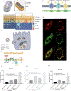
Figure 9:. Optogenetic recruitment of OCRL to the plasma membrane.
Stimulation with blue light recruits mCh-Cry2-OCRL to CIBN-CAAX-GFP localized at the plasma membrane - CIBN-GFP was modified with the targeting sequence CAAX, causing the construct to localize to the plasma membrane. Stimulation with blue light caused Cry2-OCRL-mCherry to interact with CIBN-CAAX-GFP and therefore localize to the plasma membrane. CIBN-GFP was modified with the cilia targeting sequence from the Somatostatin receptor 3 protein (SSTR3), causing the construct to localize to the plasma membrane. Stimulation with blue light caused Cry2-OCRL-mCherry to therefore localize to the primary cilia. Reprinted with author access through Translational Vision Science & Technology.
Figure 10:
Figure 10:. Trabecular meshwork targeting and functional rescue using CRY2/CIBN optogenetics
A. AAV2-s injected into the anterior chamber was used to transduce trabecular meshwork with the optogenetics CRY2/CIBN system. Plasma membrane targeting AAV2-s-CIBN-EGFP-CAAX was observed in the trabecular meshwork 4 weeks post-injection. Control trabecular meshwork did not have any GFP signal. B. Outflow facility was measured by perfusion. Blue light stimulation of the CRY2/CIBN system, which caused OCRL to localize to the plasma membrane, rescued the decreased outflow facility observed in the glaucomatous eyes of the Lowe syndrome mouse model. C. Additionally, tonometer readings indicate that the intraocular pressure was reduced with blue light stimulation. Reprinted with author access through Translational Vision Science & Technology. D. AAV2-s injected into the anterior chamber was used to transduce trabecular meshwork with the optogenetics CRY2/CIBN system. Plasma membrane targeting AAV2-s-CIBN-EGFP-CAAX was observed in the trabecular meshwork 4 weeks post-injection. Control trabecular meshwork did not have any GFP signal. E. Outflow facility was measured by perfusion. Blue light stimulation of the CRY2/CIBN system, which caused OCRL to localize to the plasma membrane, rescued the decreased outflow facility observed in the glaucomatous eyes of the Lowe syndrome mouse model. F. Additionally, tonometer readings indicate that the intraocular pressure was reduced with blue light stimulation. Reprinted with author access through Translational Vision Science & Technology.
Figure 11:
Figure 11:. MTP18 a gene known that promotes mitochondrial fragmentation was knocked down to promote neurite outgrowth on inhibitory substrate but is not sufficient for promoting axon regeneration after optic nerve crush injury.
A. Representative images of cultured RGCs with neurite growth shown after electroporation with a non-targeting scramble siRNA or MTP18 targeting siRNA and seeding on PDL + Laminin or PDL + Laminin + CSPGs (3 μg/ml) for 72 hrs (scale bar 100 μm). B. Average neurite length of cultured RGCs electroporated with scramble siRNA or MTP18 siRNA and seeded onto different concentrations of CSPGs. All points are N = 4 repeat electroporation experiments per point, normalized to average neurite length of scramble siRNA seeded onto PDL + Laminin only treated wells (significance between scramble and MTP18 siRNA treatments at each CSPG concentration was determined by Student’s t-test, *p ≤ 0.05). C. Scrambled and MTP18 siRNA-electroporated RGCs were plated at the same density as in neurite outgrowth experiments and were assayed for living and dead cells, using calcein AM (green labeled live cells) and sytox (orange labeled dead cells). Tiled images were captured per well and green and red cells were counted and graphed as percent ratio, live over dead cells, normalized to scrambled siRNA controls. (N = 4 repeat electroporation experiments, significance tested for by Student’s t-test, p ≥ 0.05.) D. Representative fluorescent images of P40 rats intravitreally injected with AAV2 viruses expressing shRNA against MTP18 (Anti-MTP18) or control shRNA against a non-present luciferase gene (Anti-luciferase) prior to optic nerve crush. CTB Alexa-555 labeled axons after optic nerves crush show no significant regeneration. Mitochondrial (mito.) mTurquoise labeling from prior virus transduction and merged images with CTB, show degenerate labeling past the crush site and bright linear labeling of some preserved axons, just prior to the crush site (left side of images, scale bar 500 μm). Reprinted with open access and approval through creative commons (creativecommons.org)
Figure 12:
Figure 12:. ORCA Schematic and rationale.
A. Model for the ORCA approach: inserting type 1 proton (H+) pumping opsins into the inner mitochondrial membrane will allow ocular cells exposed to light to generate a mitochondrial H+ gradient for subsequent ATP production via ATP synthase but independent of the rest of the electron transport chain (ETC) machinery. B. To test this hypothesis, we selected three different H+ pumping opsins not previously tested in the ophthalmology field. bR, dR, and xR generalized structures shown, along with activation wavelengths. C. Human retinal pigment epithelial (hRPE) cells were transfected with each plasmid using lipofectamine. The plasmid sat extrachromosomally in the nucleus, read by host hRPE machinery, and shuttled to the inner mitochondrial membrane D. Transfected cells show that opsins colocalize with mitochondria. Immunofluorescent staining showed opsins (red), detected by anti-flag antibody (F1804, Sigma-Aldrich) and Alexa Fluor Plus 555 (A32727, ThermoFisher Sci.), colocalize with mitochondria (green), in cells transfected with a plasmid encoding modified opsins and a mitochondrial targeted GFP. E. We selected toxins that inhibit the mitochondrial electron transport chain (ETC) to simulate clinical mitochondrial dysfunction. Rotenone inhibits Complex I, decreasing the buildup of a transmembrane proton gradient. F. hRPE plated on 96 well plates were transfected with modified opsins or empty plasmids (control plasmid), exposed to 1 uM Rotenone for 24hrs, and then exposed to 1 hr of light stimulation. ATP levels where then detected on a plate reader using a luminescence assay (CellTiter-Glo® 2.0, G9241, Promega), according to manufacturer specifications. Comparing LS_bR Plasmid to LS_Control Plasmid suggests that the H+ opsin bacteriorhodopsin (bR) may have an effect in increasing hRPE ATP in the presence of rotenone. G. A similar trend was observed for deltarhodopsin (dR). H. A similar trend was observed for xanthorhodopsin (xR).

Similar articles

  • Advances in Ophthalmic Optogenetics: Approaches and Applications.
    Prosseda PP, Tran M, Kowal T, Wang B, Sun Y. Prosseda PP, et al. Biomolecules. 2022 Feb 8;12(2):269. doi: 10.3390/biom12020269. Biomolecules. 2022. PMID: 35204770 Free PMC article. Review.
  • Opsins for vision restoration.
    Simon CJ, Sahel JA, Duebel J, Herlitze S, Dalkara D. Simon CJ, et al. Biochem Biophys Res Commun. 2020 Jun 25;527(2):325-330. doi: 10.1016/j.bbrc.2019.12.117. Epub 2020 Jan 23. Biochem Biophys Res Commun. 2020. PMID: 31982136 Review.
  • Optogenetic Strategies for Vision Restoration.
    Lu Q, Pan ZH. Lu Q, et al. Adv Exp Med Biol. 2021;1293:545-555. doi: 10.1007/978-981-15-8763-4_38. Adv Exp Med Biol. 2021. PMID: 33398841 Review.
  • Optogenetics.
    Duebel J, Marazova K, Sahel JA. Duebel J, et al. Curr Opin Ophthalmol. 2015 May;26(3):226-32. doi: 10.1097/ICU.0000000000000140. Curr Opin Ophthalmol. 2015. PMID: 25759964 Free PMC article. Review.
  • Optogenetic Therapy for Visual Restoration.
    Sakai D, Tomita H, Maeda A. Sakai D, et al. Int J Mol Sci. 2022 Nov 30;23(23):15041. doi: 10.3390/ijms232315041. Int J Mol Sci. 2022. PMID: 36499371 Free PMC article. Review.

Cited by

References

    1. Adamantidis A, Arber S, Bains JS, Bamberg E, Bonci A, Buzsáki G, Cardin JA, Costa RM, Dan Y, Goda Y, Graybiel AM, Häusser M, Hegemann P, Huguenard JR, Insel TR, Janak PH, Johnston D, Josselyn SA, Koch C, Kreitzer AC, Lüscher C, Malenka RC, Miesenböck G, Nagel G, Roska B, Schnitzer MJ, Shenoy KV, Soltesz I, Sternson SM, Tsien RW, Tsien RY, Turrigiano GG, Tye KM, Wilson RI, 2015. Optogenetics: 10 years after ChR2 in neurons--views from the community. Nat. Neurosci. 18, 1202–1212. - PubMed
    1. Aisenbrey S, Lafaut BA, Szurman P, 2002. Macular translocation with 360 retinotomy for exudative age-related macular degeneration. Archives of. - PubMed
    1. Alam NM, Mills WC 4th, Wong AA, Douglas RM, Szeto HH, Prusky GT, 2015. A mitochondrial therapeutic reverses visual decline in mouse models of diabetes. Dis. Model. Mech. 8, 701–710. - PMC - PubMed
    1. Alamusi Matsuo, T., Hosoya O, Uchida T, 2017. Visual evoked potential in RCS rats with Okayama University-type retinal prosthesis (OUReP) implantation. J. Artif. Organs 20, 158–165. - PMC - PubMed
    1. Arcuri J, Liu Y, Lee RK, Bhattacharya SK, 2020. Lipid profile dataset of optogenetics induced optic nerve regeneration. Data Brief 31, 106001. - PMC - PubMed

Publication types

LinkOut - more resources