Review
. 2023 Jan:192:114635.
doi: 10.1016/j.addr.2022.114635. Epub 2022 Nov 26.

Protein corona: Friend or foe? Co-opting serum proteins for nanoparticle delivery

Affiliations
Review

Protein corona: Friend or foe? Co-opting serum proteins for nanoparticle delivery

Woojun Kim et al. Adv Drug Deliv Rev. 2023 Jan.

Abstract

For systemically delivered nanoparticles to reach target tissues, they must first circulate long enough to reach the target and extravasate there. A challenge is that the particles end up engaging with serum proteins and undergo immune cell recognition and premature clearance. The serum protein binding, also known as protein corona formation, is difficult to prevent, even with artificial protection via "stealth" coating. Protein corona may be problematic as it can interfere with the interaction of targeting ligands with tissue-specific receptors and abrogate the so-called active targeting process, hence, the efficiency of drug delivery. However, recent studies show that serum protein binding to circulating nanoparticles may be actively exploited to enhance their downstream delivery. This review summarizes known issues of protein corona and traditional strategies to control the corona, such as avoiding or overriding its formation, as well as emerging efforts to enhance drug delivery to target organs via nanoparticles. It concludes with a discussion of prevailing challenges in exploiting protein corona for nanoparticle development.

Keywords: Drug delivery; Nanoparticles; Protein corona; Stealth; Targeting.


Conflict of interest statement

Declaration of Competing Interest The authors declare that they have no known competing financial interests or personal relationships that could have appeared to influence the work reported in this paper.

Figures

Figure 1.
Figure 1.
(a) Different methods to coat PLGA NPs with albumin. NP×Al: interfacial embedding; NP-pD-Al: polydopamine-mediated physisorption. (b) Top: Representative SDS-PAGE gel image of albumin after pulse proteolysis. Native albumin (nAlb), denatured albumin (dAlb), NP×Al, and NP-pD-Al were treated with thermolysin for 3 min. Lane 1: nAlb; Lane 2: dAlb; Lane 3: NP×Al; Lane 4: NP-pD-Al; Lane 5: nAlb + thermolysin; Lane 6: dAlb + thermolysin; Lane 7: NP×Al + thermolysin; and Lane 8: NP-pD-Al + thermolysin. Bottom: % digestion albumin was defined as (1-albumin band intensity after proteolysis/albumin band intensity prior to proteolysis) × 100. (c) Schematic of a Transwell co-culture system with HUVEC in the insert and B16F10 cells in the bottom of the basolateral side (left top); Transendothelial electrical resistance (TEER) indicating the confluence of HUVEC layer at the time of NP application (left bottom); NP associated with B16F10 cells, measured at 24 h after 6 h incubation with a Transwell containing NPs and the confluent HUVEC layer. (d) Dosing schedule of PTX-loaded NPs (top); PTX content in B16F10 tumors treated with PTX-loaded NP×Al (PTX@NP×Al) or PTX-loaded NP-pD-Al (PTX@NP-pD-Al) (bottom left); % injected PTX dose per gram of each tissues (%ID/g) of PTX@NP×Al or PTX@NP-pD-Al in B16F10 tumor bearing mice 24 h after i.v. injection. %ID/g is defined as percentage of injected dose per gram of tissue weight. Reprinted from [161] with permission.
Figure 2.
Figure 2.
(a) Schematic illustration of Nanosac (MSNa/siRNA/pD) preparation and transmission electron microscopy images of Nanosac and Nanosac precursors. Nanosac is produced by sequential coating of mesoporous silica nanoparticles (MSNs) with siRNA and polydopamine, followed by removal of the sacrificial MSN core. (b) Confocal microscope images of cy5-labeled MSNa, MSNa/pD, and Nanosac relative to lysosomes in CT26 cells and fluorescence intensity profiles along the white lines in confocal images. Green: Lysotracker (lysosome); red: cy5- labeled NPs; and blue: Hoechst 33342 (nuclei). Scale bars: 10 μm. (c) SDS-PAGE of protein corona composition formed on MSNa, MSNa/pD, and Nanosac. The protein corona bound on each NPs were further analyzed by LC-MS/MS for most abundant proteins. 4 mg/mL of each NPs were incubated in 50% FBS for 2 h and rinsed with PBS twice. (d) Average tumor size after treatment of anti-PD-L1 antibody and siPD-L1-Nanosac to Balb/c mice bearing CT26 tumors. (anti-PD-L1 antibody: 200 μg/mouse/time, intraperitoneal injection; siPD-L1:1.5 mg/kg/time, IV injection; q2d × 5). Reprinted from [55] with permission.
Figure 3.
Figure 3.
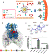
(a) Schematics Illustration of gold NPs targeting TfR overexpressed on cancer cells. (b) Five potential binding sites, Pocket 1 through 5, identified on the human Tf predicted by Fpocket. Tf is represented with the blue-ribbon diagram, and part of the ectodomain of the transferrin receptor dimer23 is rendered by its accessible surface. Due to the adequate volume for peptide and the distance from transferrin or iron binding site, Pocket 3 (orange) is chosen to dock the peptide. (c) 3D docked pose of the synthesized Tf-binding peptide (Tf2) created by coarse-grained molecular dynamics simulation. Atoms are colored according to their root mean squared displacement. Blue: rigid regions; red: flexible regions; green dashed lines: hydrogen bonds; red dashed lines: salt bridges; black dashed lines: solvent exposed atoms. (d) Cell uptake of gold NPs conjugated with Tf2. Various gold NPs were prepared using different percentage of PepN-Tf2 (0, 1, and 10% w/w). NPs were incubated in human plasma for 1 h at 37 °C and then with Mia PaCa-2 cells for 1 h at 37 °C in DMEM with (right, suffix +Tf2) or without (left) a Tf2. The results were normalized to the amount of internalized gold in AuNP-0. Reprinted from [201] with permission.
Figure 4.
Figure 4.
Tf targeting in glioma by DOX-loaded, T10-coated COF NPs (DCPT) NPs. (a) Schematic illustration of endogenous Tf corona-mediated DCPT delivery across the BBB. (b) SDS-PAGE analysis of PC on DCPT-2 before and after passage through the in vitro BBB model (left). FBS-Tf: Formation of Tf corona on the surface of DCPT-2 mediated by Tf from the FBS. SD rat-Tf: Formation of Tf corona on the surface of DCPT-2 mediated by Tf from the SD rat serum. Cellular uptake of DOX and COF formulations incubated with U87 cells under different conditions (right). (c) Ex vivo imaging of DOX in main organs of glioma-bearing mice after intravenous injection of DOX, Caelyx, DCP (DOX-loaded COF, no T10) and DCPT-2 at 12 h (left). Immunofluorescence images of brain sections from orthotopic glioma mice after 12 h post-injection of DCP, Caelyx and DCPT-2, respectively. Blue: nuclei; purple: U87 cells; red: DOX; green: anti-CD31 labeled blood vessels. White arrows: co-localization of DOX and blood vessels; Yellow arrows: co-localization of DOX and glioma cells. Bar: 200 μm. Reprinted from [205] with permission.
Figure 5.
Figure 5.
Schematic illustrations of (a) differential organ distribution of O- and N-series LNPs and (b) interaction of LNPs with proteins in the blood vessel. Quantification of the percentage of total proteins of the top three protein components in the protein corona of (c) the O-series LNPs (306-O12B) and (d) the N-series LNPs (306-N16B). Top 20 most abundant corona proteins based on (e) their calculated molecular weight, (f) isoelectric point, and (g) biological function. Reprinted from [215] with permission.

Similar articles

Cited by

References

    1. Matsumura Y, Maeda H, A New Concept for Macromolecular Therapeutics in Cancer Chemotherapy: Mechanism of Tumoritropic Accumulation of Proteins and the Antitumor Agent Smancs, Cancer Res, 46 (1986) 6387–6392. - PubMed
    1. Singh AK, Chapter 6 - Nanoparticle Pharmacokinetics and Toxicokinetics, in: Singh AK (Ed.) Engineered Nanoparticles, Academic Press, Boston, 2016, pp. 229–293.
    1. Zhang H, Burnum KE, Luna ML, Petritis BO, Kim J-S, Qian W-J, Moore RJ, Heredia-Langner A, Webb-Robertson B-JM, Thrall BD, Camp DG, Smith RD, Pounds JG, Liu T, Quantitative proteomics analysis of adsorbed plasma proteins classifies nanoparticles with different surface properties and size, Proteomics, 11 (2011) 4569–4577. - PMC - PubMed
    1. Monopoli MP, Aberg C, Salvati A, Dawson KA, Biomolecular coronas provide the biological identity of nanosized materials, Nat Nanotechnol, 7 (2012) 779–786. - PubMed
    1. Sepand MR, Ghavami M, Zanganeh S, Stacks S, Ghasemi F, Montazeri H, Corbo C, Derakhshankhah H, Ostad SN, Ghahremani MH, Mahmoudi M, Impact of plasma concentration of transferrin on targeting capacity of nanoparticles, Nanoscale, 12 (2020) 4935–4944. - PubMed

Publication types