Review
. 2022 Dec 1:18:100508.
doi: 10.1016/j.mtbio.2022.100508. eCollection 2023 Feb.

Recent advances in responsive hydrogels for diabetic wound healing

Affiliations
Review

Recent advances in responsive hydrogels for diabetic wound healing

Siming Zhang et al. Mater Today Bio. .

Abstract

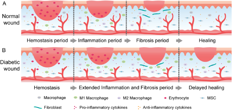
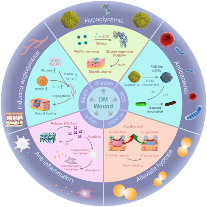
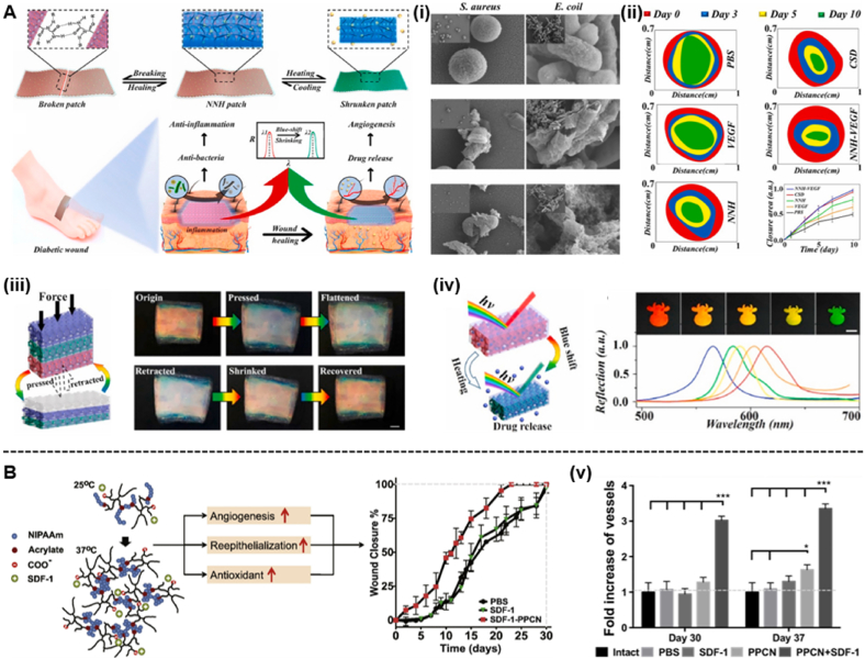
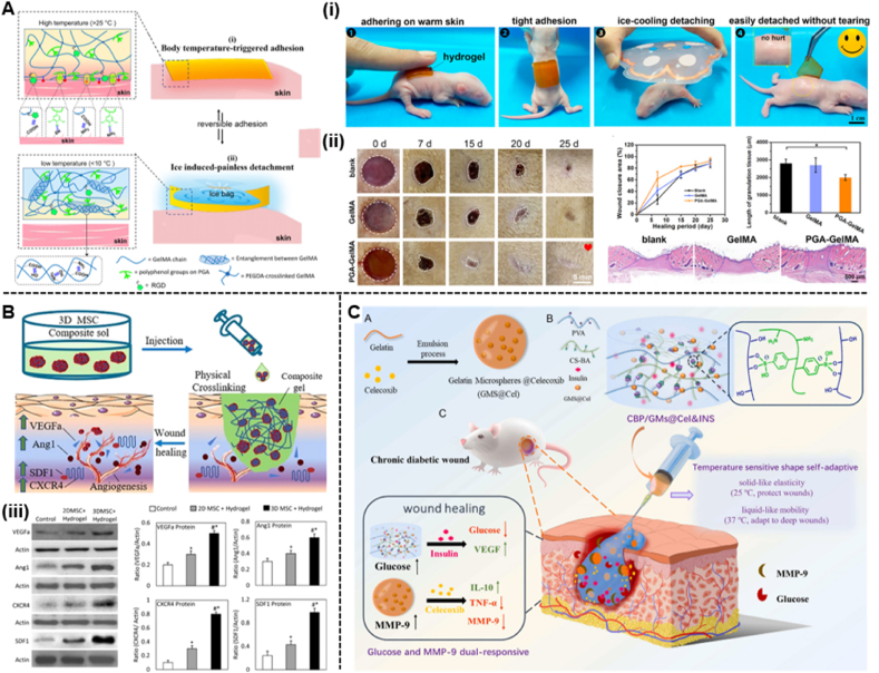
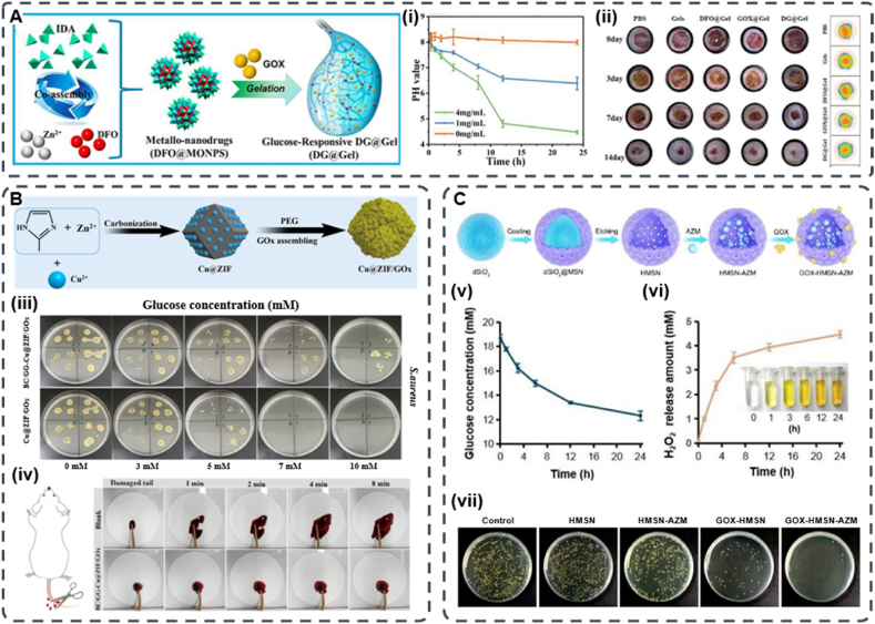
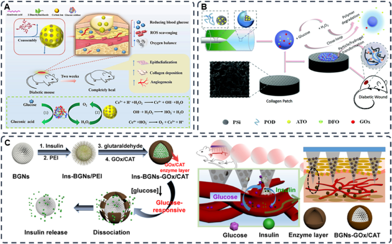
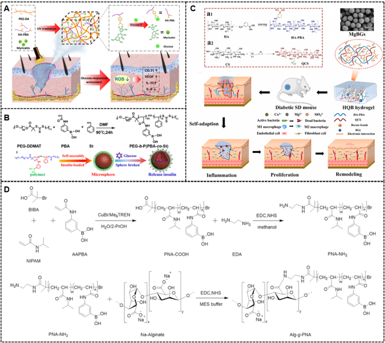
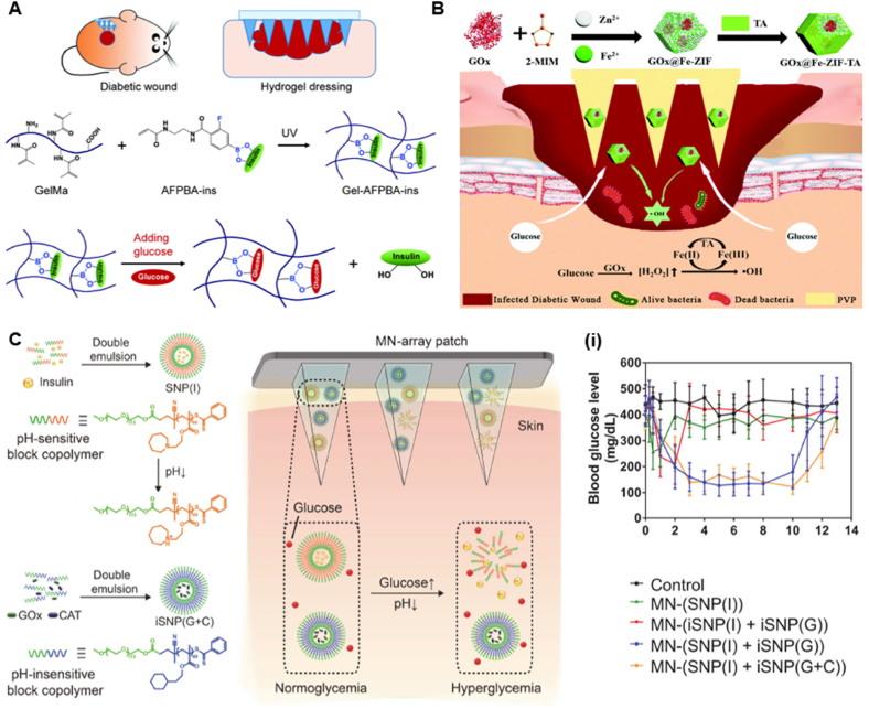
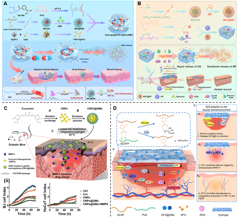
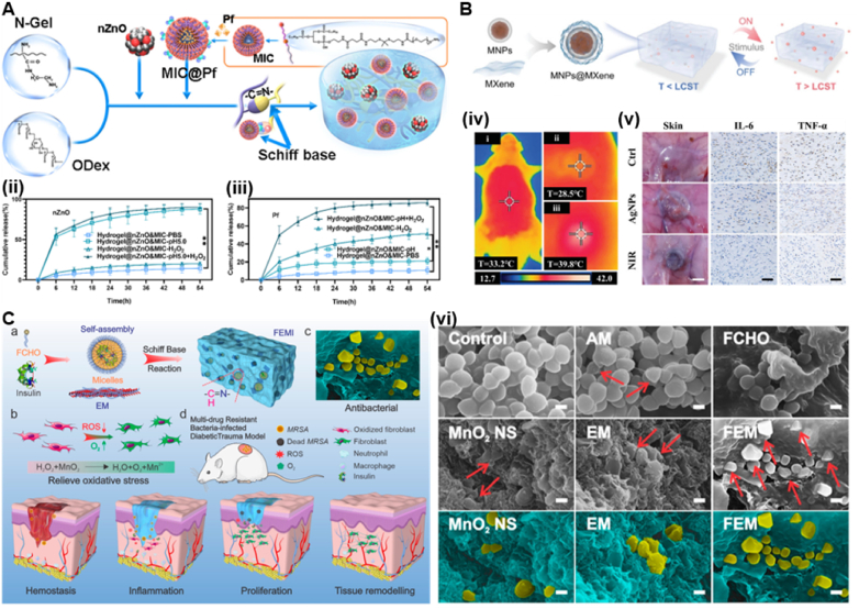
Poor wound healing after diabetes mellitus remains a challenging problem, and its pathophysiological mechanisms have not yet been fully elucidated. Persistent bleeding, disturbed regulation of inflammation, blocked cell proliferation, susceptible infection and impaired tissue remodeling are the main features of diabetic wound healing. Conventional wound dressings, including gauze, films and bandages, have a limited function. They generally act as physical barriers and absorbers of exudates, which fail to meet the requirements of the whol diabetic wound healing process. Wounds in diabetic patients typically heal slowly and are susceptible to infection due to hyperglycemia within the wound bed. Once bacterial cells develop into biofilms, diabetic wounds will exhibit robust drug resistance. Recently, the application of stimuli-responsive hydrogels, also known as "smart hydrogels", for diabetic wound healing has attracted particular attention. The basic feature of this system is its capacities to change mechanical properties, swelling ability, hydrophilicity, permeability of biologically active molecules, etc., in response to various stimuli, including temperature, potential of hydrogen (pH), protease and other biological factors. Smart hydrogels can improve therapeutic efficacy and limit total toxicity according to the characteristics of diabetic wounds. In this review, we summarized the mechanism and application of stimuli-responsive hydrogels for diabetic wound healing. It is hoped that this work will provide some inspiration and suggestions for research in this field.

Keywords: Biomaterials; Diabetic wound; Hydrogel; Skin tissue engineering; Smart responsiveness.


Conflict of interest statement

The authors declare that they have no known competing financial interests or personal relationships that could have appeared to influence the work reported in this paper.

Figures

Image 1
Graphical abstract
Fig. 1
Fig. 1
Schematic representation of the main stages of normal wound healing (A) and delayed healing of diabetic wounds (B).
Fig. 2
Fig. 2
Typical alterations in diabetic wounds and a summary of the classification and biological functions of smart hydrogels.
Fig. 3
Fig. 3
Differences between the normal wound microenvironment and the diabetic wound microenvironment.
Fig. 4
Fig. 4
(A)The primary hypothesis of this study. Protamine NPs and HAO were loaded into the pores of Ca-alginate hydrogel and showed complete and fast release at the alkaline diabetic wound site. Protamine NPs/HAO Ca-alginate hydrogel was placed on the wound in streptozotocin-induced type 1 diabetic rats, and protamine NPs exhibited antibacterial activity by damaging the cell wall, thus reducing the inflammatory response. Moreover, HAO significantly enhanced HUVEC migration and capillary-like tube formation with HAO-induced stimulation of VEGF, leading to rapid diabetic wound healing. (i) Schematic representation of protaimine NPs/HAO CaAlg hydrogel. TEM image of protamine NPs and DLS analysis of the obtained protamine NPs. SEM image of protamine NPs/HAO CaAlg hydrogel and zeta potential distribution of protamine NPs. (ii) SEM images of bacteria treated with protamine NPs/HAO CaAlg hydrogel. (iii) VEGF secretion by HUVECs treated with different kinds of CaAlg hydrogels by ELISA. (iv) H&E staining of tissue sections treated with PBS, CaAlg hydrogel, and protaimine NPs/HAO CaAlg hydrogel on days 1, 3, 7, and 14 (Reproduced with permission of Ref. [89]). (B)Schematic illustration of Gel@fMLP/SiO2-FasL for transiently heightened inflammatory response to initiate refractory wound healing. SiO2-FasL, silicon dioxide nanoparticles conjugated with FasL; Gel@fMLP/SiO2-FasL, pH-responsive hydrogel matrix loaded with fMLP and SiO2-FasL. (v) IR spectrometry of CS-FPBA, fMLP and Gel@fMLP/SiO2-FasL. Photographs of the gelation process of Gel@fMLP/SiO2-FasL with a solution containing RhB (representing fMLP) and SiO2-FasL after incubation at pH 7.4. The decomposition of Gel@fMLP/SiO2-FasL at pH 5.5. (vi) Critical-sized calvarial bone defects were generated in 8-week-old female C57BL/6 mice and treated with hydrogels hybridized with different components. The mice were sacrificed at different time points post operation for further examination. Micro-CT analysis of bone defects (Reproduced with permission of Ref. [91]).
Fig. 5
Fig. 5
(A) Synthesis of oxidized hyaluronic acid (HA); Schiff base reaction between oxidized HA and polypeptide (ε-poly-l-lysine, EPL); thermal-responsive sol-gel process of double network hydrogel composed of F127-EPL and oxidized HA. (i) pH-dependent release profile of loaded exosomes in the FHE hydrogel. (ii) Representative images of the healing process in wounds treated with FHE, exosomes, FHE@exosome and control (Reproduced with permission of Ref. [94]). (B) Schematic diagram of the preparation and application of the PC/GO/Met hydrogel. Preparation of dihydrocaffeic acid and l-arginine cografting chitosan (CS-DA-LAG) and phenylboronic acid and benzaldehyde difunctionalized polyethylene glycol-co-poly (glycerol sebacic acid) (PEGS-PBA-BA) and polydopamine coated rGO (rGO@PDA). (iii) Schematic of self-healing, responsive drug release, and removability given by dual dynamic bonds: self-healing mechanism and representative pictures of PC hydrogel and release mechanism of metformin in response to pH and glucose and representative display pictures of removability. (iv) Representative HE staining results of wound tissue on days 3, 7, 14, and 21 (Reproduced with permission of Ref. [95]). (C)The sketch map of “Double-H bonds” in the hydrogel. (v) Live/dead staining of NIH-3T3 cells (scale bar ​= ​200 ​mm, green – live cells, and red – dead cells) and the cytoskeleton staining of NIH-3T3 cells 3D cultured in hydrogels. (vi) Drug release curves of Met from HA–COL-GMs. (vii) Migrated fibroblast staining and collagen secretion staining (Reproduced with permission of Ref. [98]).
Fig. 6
Fig. 6
(A) Schematic representation of the excellent performance of the NNH patches and their application to diabetic wounds. (i) Images of NNH patches against S. aureus and E. coli from scanning electron microscope (SEM). (ii) In vivo diabetic wound healing. The size and quantification of the wound closure area from the PBS group, VEGF group, NNH patch group, CSD group and NNH-VEGF patch group. (iii) Scheme of the compression of NNH bulk. Representative of compression and retraction of NNH bulk. (iv) Scheme of the temperature-responsive feature of the NNH patches. Reflection images and spectra of NNH patches at a temperature of 37 ​°C (Reproduced with permission of Ref. [123]). (B) Scheme of the antioxidant thermoresponsive hydrogel. (v) Quantification of the functional blood vessel density (Reproduced with permission of Ref. [127]).
Fig. 7
Fig. 7
(A) Schematic illustrations of the body temperature-triggered gentle adhesion and ice-cooling-induced painless detachment of the PGAGelMA hydrogel. (i) Photographs of adhering and triggerable removal of the PGA-GelMA hydrogel on an infant rat skin surface. (ii) Digital images of the wound healing process in all groups and quantification of the wound closure area at different time intervals. Representative H&E staining images of wound samples in different groups on Day 25 and quantification of the length of granulation tissue (Reproduced with permission of Ref. [131]). (B) Schematic illustrations of the thermosensitive Injectable Chitosan/Collagen/β-Glycerophosphate Composite Hydrogels. (iii) Expression of paracrine cytokines in wound tissues (Reproduced with permission of Ref. [132]). (C) The design strategy of glucose and MMP-9 dual-responsive shape self-adaptive hydrogels for treating chronic diabetic wound (Reproduced with permission of Ref. [139]).
Fig. 8
Fig. 8
(A) Schematic of the preparation of DG@Gel. (i) Change in the pH of DG@Gel over 24 ​h in different glucose concentrations. (ii) Photographs of wound tissues treated with different sample groups on days 0, 3, 7, and 14, and wound healing boundaries in different treatment groups in vivo (Reproduced with permission of Ref. [144]). (B) Schematic illustration of preparing procedure of Cu@ZIF and Cu@ZIF/GOx. (iii) Antibacterial efficacy of Cu@ZIF/GOx treated with different concentration of glucose after 6 ​h incubation (Reproduced with permission of Ref. [148]). (C) Schematic illustration of the synthetic route of GOx-HMSN-AZM. (v) He glucose concentration variation under the catalyst of 200 ​μg/mL GOx-HMSN-AZM on different reaction times. (vi) The concentration of generated H2O2 catalyzed by 200 ​μg/mL GOx-HMSN-AZM on different reaction times. (vii) Photographs of bacterial colonies formed by S. aureus in biofilms which were treated with 500 ​μg/mL of PBS, HMSN, HMSN-AZM, GOx-HMSN and GOx-HMSN-AZM (Reproduced with permission of Ref. [149]).
Fig. 9
Fig. 9
(A) Schematic diagram of Ce-driven coassembly multi-enzymatic activity of nanozyme for diabetic wound healing (Reproduced with permission of Ref. [155]). (B) Schematic illustration of the construction of glucose responsive delivery system and the orchestrated cascade for diabetic wound care. The detrimental side product of H2O2, was utilized and consumed, further aiding the close-loop system (Reproduced with permission of Ref. [156]). (C) Schematic representation of the glucose-sensitive insulin delivery system using glucose-sensitive BGNs; after the treatment with diabetic rats, the glucose-sensitive insulin released from the MNs in vivo (Reproduced with permission of Ref. [158]).
Fig. 10
Fig. 10
(A) Diagram illustrating the PHM Hydrogel Platform for Diabetic Wound Healing (Reproduced with permission of Ref. [167]). (B) The synthetic route of PEG-b-P(PBA-co-St), self-assembly of PEG-b-P(PBA-co-St) micelles and insulin release from these micelles in the presence of glucose (Reproduced with permission of Ref. [169]). (C) The fabrication schematic and mechanism of a bioactive HQB hydrogel and its application on chronic wound healing (Reproduced with permission of Ref. [176]). (D) Synthetic Routes of PNA-NH2 and Alg-g-PNA. (b) Copolymers (Reproduced with permission of Ref. [181]).
Fig. 11
Fig. 11
(A) Responsive microneedle dressing for diabetic wound healing. Diabetic wounds in mice treated with the hydrogel-based microneedle dressing. Preparation of glucose-responsive insulin-releasing Gel–AFPBA–ins hydrogels and mechanism of glucose-responsive insulin release from the prepared hydrogels (Reproduced with permission of Ref. [191]). (B) Schematic illustration of preparation of hierarchically porous glucose-responsive antibacterial GOx@Fe-ZIF-TA and penetration of MOF-based MNs into infected diabetic wounds to release antibacterial particles for infected diabetic wound treatment (Reproduced with permission of Ref. [193]). (C) Scheme of MN-array patch integrated with pH-sensitive insulin nanoformulations for glucose-responsive insulin delivery. The chemical structure and formation of SNP(I) and iSNP(G ​+ ​C) and schematic illustration of the MN-array patch loaded with SNP(I) and iSNP(G ​+ ​C) for in vivo insulin delivery triggered by a hyperglycemic state to release more insulin. (i) Blood glucose levels of type 1 diabetic mice after treatment with blank MNs as control, MNs loaded with SNP(I), MNs loaded with iSNP(I) and iSNP(G), MNs loaded with SNP(I) and iSNP(G), MNs loaded with SNP(I) and iSNP(G ​+ ​C) (Reproduced with permission of Ref. [198]).
Fig. 12
Fig. 12
(A) Diagrams displaying the formation of NIM-Loaded Micelle, VAN-AgNCs, Hydrogels, and the mechanisms of accelerating wound healing (Reproduced with permission of Ref. [212]). (B) Schematic diagram of the fabrication procedures of the DS&MIC@MF embedded POD/CE hydrogels and illustration of spatiotemporally drugs release behaviour of the hydrogel, and the mechanism of the hydrogel for accelerating wound healing on the infected diabetic cutaneous wound model (Reproduced with permission of Ref. [214]). (C) Schematic representations of CNPs@GMs/hydrogel preparation and the process of drug release at the wound bed in diabetic mice. Preparation of pure CNPs via a solution exchange method. CNPs loaded into GMs by the emulsion process to get CNPs@GMs. CNPs@GMs mixed with the thermos-sensitive hydrogel and covered on the wound in diabetic mice. Under the microenvironment of a nonhealing wound, GMs were degraded by MMPs, and specifically the drug was specifically released. (ii) Cell migration/invasion in real time was monitored by the xCELLigence RTCA for 72 ​h (Reproduced with permission of Ref. [223]). (D) The fabrication and application of self-adaptive DFO@G-QCSFP for accelerating diabetic wound healing on the full-thickness diabetic wound of a diabetic SD rat. a) The chemical structure of the hydrogel and the mechanism of the hydrogel for accelerating diabetic wound healing. b) The self-adaption of the hydrogel to wound microenvironment. The hydrogel reduced oxidative stress and released DFO@G on demand. Then DFO was released and promoted angiogenesis (Reproduced with permission of Ref. [224]).
Fig. 13
Fig. 13
(A) Illustration of microenvironment-Responsive Hydrogel fabrication stragety. (ii) The release tendency of nZnO and Pf (iii) over 54 ​h (Reproduced with permission of Ref. [226]). (B) Schematic illustration of the preparation of stimuli-responsive MXene-based hydrogel system. The formation and drug release process of the MXene-based hydrogel system. (iv) Thermal images of AgNPs-loaded MXene-based hydrogel applied to the rat subcutaneous before and after near-infrared irradiation. (v) Photos of the subcutaneous tissue of the diabetic rats and immunostaining of IL-6 and TNF-α (Reproduced with permission of Ref. [231]). (C) Schematic diagram illustrating FEMI hydrogel for MDR bacteria-infected diabetic wound healing. (vi) Representative SEM images of MRSA cells after different treatments. The yellow pseudocolor indicates the MRSA bacterial cells and blue pseudocolor indicates the surrounding materials (Reproduced with permission of Ref. [227]).

References

    1. Shaw J.E., Sicree R.A., Zimmet P.Z. Global estimates of the prevalence of diabetes for 2010 and 2030. Diabetes Res. Clin. Pract. 2010;87(1):4–14. doi: 10.1016/j.diabres.2009.10.007. - DOI - PubMed
    1. Whiting D.R., Guariguata L., Weil C., Shaw J. IDF diabetes atlas: global estimates of the prevalence of diabetes for 2011 and 2030. Diabetes Res. Clin. Pract. 2011;94(3):311–321. doi: 10.1016/j.diabres.2011.10.029. - DOI - PubMed
    1. Patel S., Srivastava S., Singh M.R., Singh D. Mechanistic insight into diabetic wounds: pathogenesis, molecular targets and treatment strategies to pace wound healing. Biomed. Pharmacother. 2019;112 doi: 10.1016/j.biopha.2019.108615. - DOI - PubMed
    1. Mogoşanu G.D., Grumezescu A.M. Natural and synthetic polymers for wounds and burns dressing. Int. J. Pharm. 2014;463(2):127–136. doi: 10.1016/j.ijpharm.2013.12.015. - DOI - PubMed
    1. Eleftheriadou I., Tentolouris N., Argiana V., Jude E., Boulton A.J. Methicillin-resistant Staphylococcus aureus in diabetic foot infections. Drugs. 2010;70(14):1785–1797. doi: 10.2165/11538070-000000000-00000. - DOI - PubMed

LinkOut - more resources