Dysphagia assessment in ischemic stroke after mechanical thrombectomy: When and how?
- PMID: 36504648
- PMCID: PMC9726734
- DOI: 10.3389/fneur.2022.1024531
Dysphagia assessment in ischemic stroke after mechanical thrombectomy: When and how?
Abstract
Background: Dysphagia is a frequent symptom in acute ischemic stroke (AIS). Endovascular treatment (EVT) has become the standard of care for acute stroke secondary to large vessel occlusion. Although standardized guidelines for poststroke dysphagia (PSD) management exist, they do not account for this setting in which patients receive EVT under general anesthesia. Therefore, the aim of this study was to evaluate PSD prevalence and severity, as well as an appropriate time point for the PSD evaluation, in patients undergoing EVT under general anesthesia (GA).
Methods: We prospectively included 54 AIS patients undergoing EVT under GA. Fiberoptic Endoscopic Evaluation of Swallowing (FEES) was performed within 24 h post-extubation in all patients. Patients presenting significant PSD received a second FEES-assessment to determine the course of dysphagia deficits over time. Dysphagia severity was rated according the Fiberoptic Dysphagia Severity Scale (FEDSS).
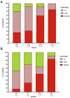
Results: At first FEES (FEES 1) assessment, performed in the median 13 h (IQR 5-17) post-extubation, 49/54 patients (90.7%) with dysphagia were observed with a median FEDSS of 4 (IQR 3-6). Severe dysphagia requiring tube feeding was identified in 28/54 (51.9%) subjects, whereas in 21 (38.9%) patients early oral diet with certain food restrictions could be initiated. In the follow up FEES examination conducted in the median 72 h (IQR 70-97 h) after initial FEES 34/49 (69.4%) patients still presented PSD. Age (p = 0.030) and ventilation time (p = 0.035) were significantly associated with the presence of PSD at the second FEES evaluation. Significant improvement of dysphagia frequency (p = 0.006) and dysphagia severity (p = 0.001) could be detected between the first and second dysphagia assessment.
Conclusions: PSD is a frequent finding both immediately within 24 h after extubation, as well as in the short-term course. In contrast to common clinical practice, to delay evaluation of swallowing for at least 24 h post-extubation, we recommend a timely assessment of swallowing function after extubation, as 50% of patients were safe to begin oral intake. Given the high amount of severe dysphagic symptoms, we strongly recommend application of instrumental swallowing diagnostics due to its higher sensitivity, when compared to clinical swallowing examination. Furthermore, advanced age, as well as prolonged intubation, were identified as significant predictors for delayed recovery of swallowing function.
Keywords: aspiration; dysphagia; endovascular treatment; fiberoptic endoscopic evaluation of swallowing (FEES); stroke; thrombectomy.
Copyright © 2022 Lapa, Neuhaus, Harborth, Neef, Steinmetz, Foerch and Reitz.
Conflict of interest statement
The authors declare that the research was conducted in the absence of any commercial or financial relationships that could be construed as a potential conflict of interest.
Figures


References
-
- Donovan NJ, Daniels SK, Edmiaston J, Weinhardt J, Summers D, Mitchell PH, et al. Dysphagia screening: state of the art: invitational conference proceeding from the state-of-the-art nursing symposium, international stroke conference 2012. Stroke. (2013) 44:e24–31. 10.1161/STR.0b013e3182877f57 - DOI - PubMed
LinkOut - more resources
Full Text Sources

