

Predicting the brain age of children with cerebral palsy using a two-dimensional convolutional neural networks prediction model without gray and white matter segmentation
- PMID: 36504669
- PMCID: PMC9730825
- DOI: 10.3389/fneur.2022.1040087
Predicting the brain age of children with cerebral palsy using a two-dimensional convolutional neural networks prediction model without gray and white matter segmentation
Abstract
Background: Abnormal brain development is common in children with cerebral palsy (CP), but there are no recent reports on the actual brain age of children with CP.
Objective: Our objective is to use the brain age prediction model to explore the law of brain development in children with CP.
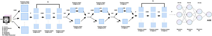
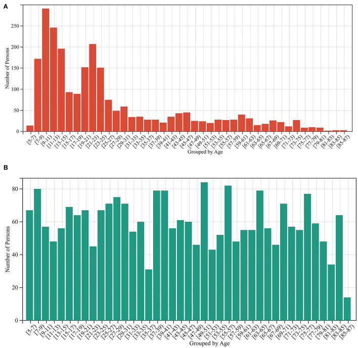
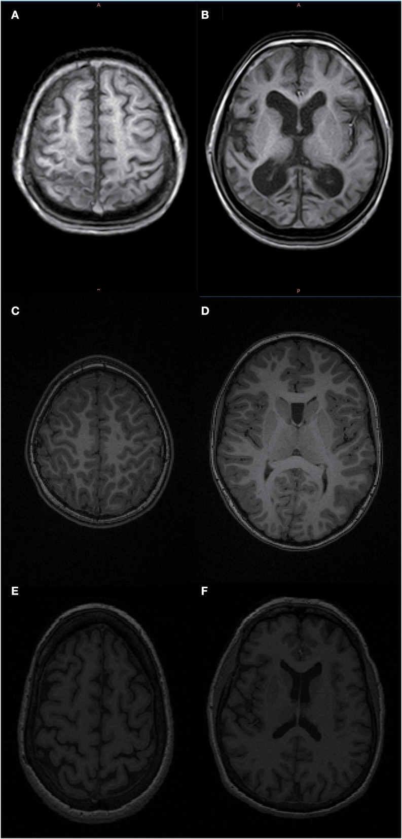
Methods: A two-dimensional convolutional neural networks brain age prediction model was designed without segmenting the white and gray matter. Training and testing brain age prediction model using magnetic resonance images of healthy people in a public database. The brain age of children with CP aged 5-27 years old was predicted.
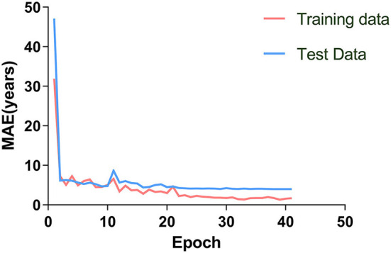
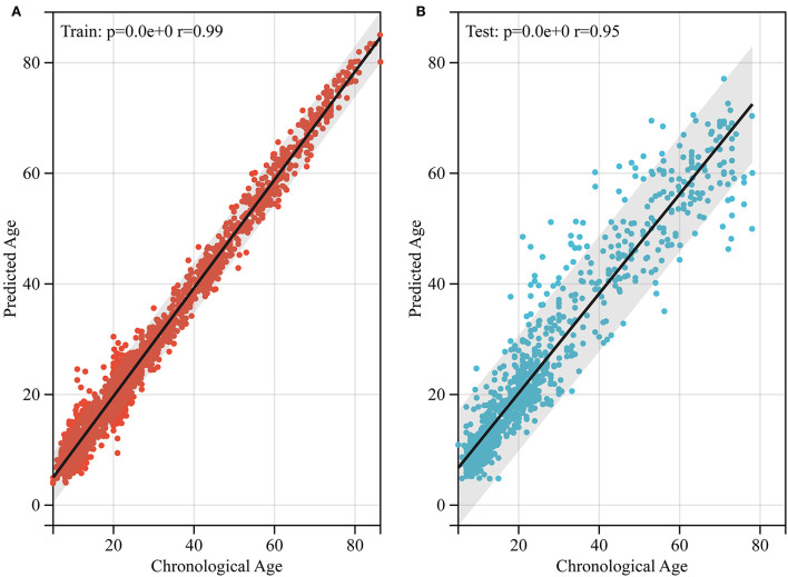
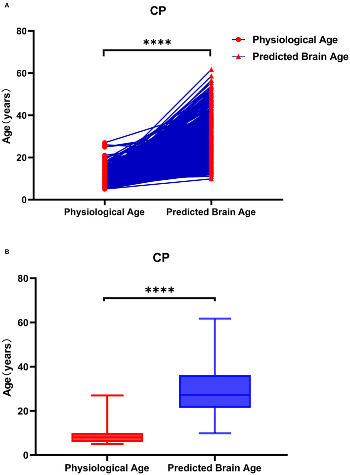
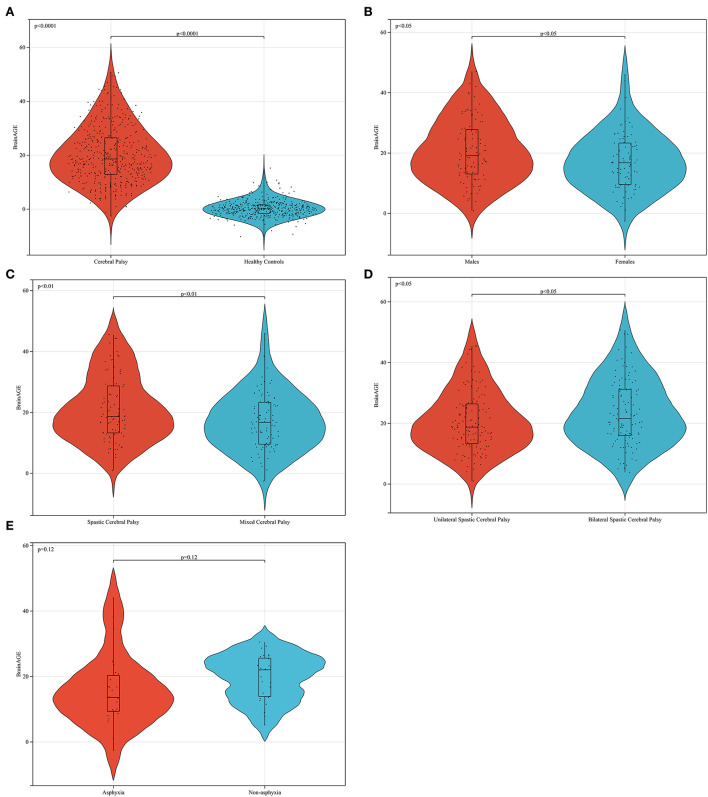
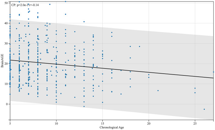
Results: The training dataset mean absolute error (MAE) = 1.85, r = 0.99; test dataset MAE = 3.98, r = 0.95. The brain age gap estimation (BrainAGE) of the 5- to 27-year-old patients with CP was generally higher than that of healthy peers (p < 0.0001). The BrainAGE of male patients with CP was higher than that of female patients (p < 0.05). The BrainAGE of patients with bilateral spastic CP was higher than those with unilateral spastic CP (p < 0.05).
Conclusion: A two-dimensional convolutional neural networks brain age prediction model allows for brain age prediction using routine hospital T1-weighted head MRI without segmenting the white and gray matter of the brain. At the same time, these findings suggest that brain aging occurs in patients with CP after brain damage. Female patients with CP are more likely to return to their original brain development trajectory than male patients after brain injury. In patients with spastic CP, brain aging is more serious in those with bilateral cerebral hemisphere injury than in those with unilateral cerebral hemisphere injury.
Keywords: brain age; brain age gap estimation; cerebral palsy; convolutional neural networks; deep learning.
Copyright © 2022 Zhang, Yan, Mutalifu, Fu, Shao, Wu, Guan, Biedelehan, Tong and Luan.
Conflict of interest statement
The authors declare that the research was conducted in the absence of any commercial or financial relationships that could be construed as a potential conflict of interest.
Figures
Similar articles
-
Assessment of the structural brain network reveals altered connectivity in children with unilateral cerebral palsy due to periventricular white matter lesions.Neuroimage Clin. 2014 Jun 4;5:84-92. doi: 10.1016/j.nicl.2014.05.018. eCollection 2014. Neuroimage Clin. 2014. PMID: 25003031 Free PMC article.
-
Brain Lesions in Children with Unilateral Spastic Cerebral Palsy.Med Arch. 2017 Feb;71(1):7-11. doi: 10.5455/medarh.2017.71.7-11. Epub 2017 Feb 5. Med Arch. 2017. PMID: 28428665 Free PMC article.
-
Athetotic and spastic cerebral palsy: anatomic characterization based on diffusion-tensor imaging.Radiology. 2011 Aug;260(2):511-20. doi: 10.1148/radiol.11101783. Epub 2011 May 9. Radiology. 2011. PMID: 21555354
-
Rodent Hypoxia-Ischemia Models for Cerebral Palsy Research: A Systematic Review.Front Neurol. 2016 Apr 25;7:57. doi: 10.3389/fneur.2016.00057. eCollection 2016. Front Neurol. 2016. PMID: 27199883 Free PMC article. Review.
-
The relationship between neuroimaging and motor outcome in children with cerebral palsy: A systematic review-Part B diffusion imaging and tractography.Res Dev Disabil. 2020 Feb;97:103569. doi: 10.1016/j.ridd.2019.103569. Epub 2019 Dec 31. Res Dev Disabil. 2020. PMID: 31901671
Cited by
-
Trends in brain MRI and CP association using deep learning.Radiol Med. 2024 Nov;129(11):1667-1681. doi: 10.1007/s11547-024-01893-w. Epub 2024 Oct 10. Radiol Med. 2024. PMID: 39388027 Free PMC article.
References
-
- Xue Y, Shi S, Zheng S, Yang Z, Xu J, Gong F. Therapeutic effect of scalp-based acupuncture and moxibustion as an adjunctive treatment on children with cerebral palsy comparing to conventional rehabilitation therapy: a systematic review and meta-analysis of randomized controlled trials. Transl Pediatr. (2022) 11:631–41. 10.21037/tp-22-85 - DOI - PMC - PubMed
LinkOut - more resources
Full Text Sources
Miscellaneous