. 2022 Nov 24:16:1067409.
doi: 10.3389/fnbeh.2022.1067409. eCollection 2022.

Loss of GABA co-transmission from cholinergic neurons impairs behaviors related to hippocampal, striatal, and medial prefrontal cortex functions

Affiliations

Loss of GABA co-transmission from cholinergic neurons impairs behaviors related to hippocampal, striatal, and medial prefrontal cortex functions

R Oliver Goral et al. Front Behav Neurosci. .

Abstract

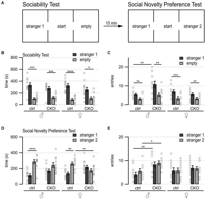
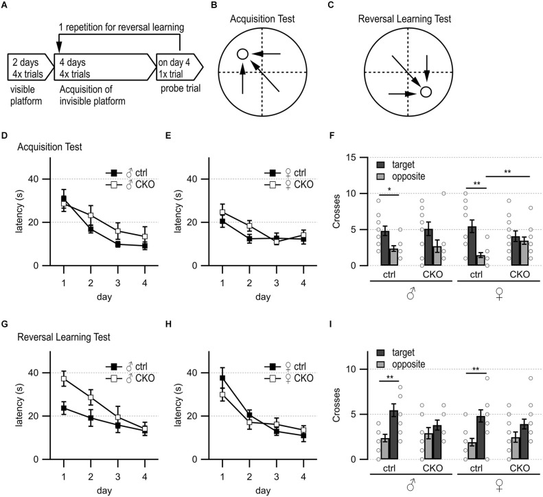
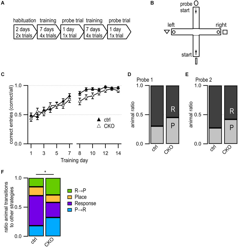
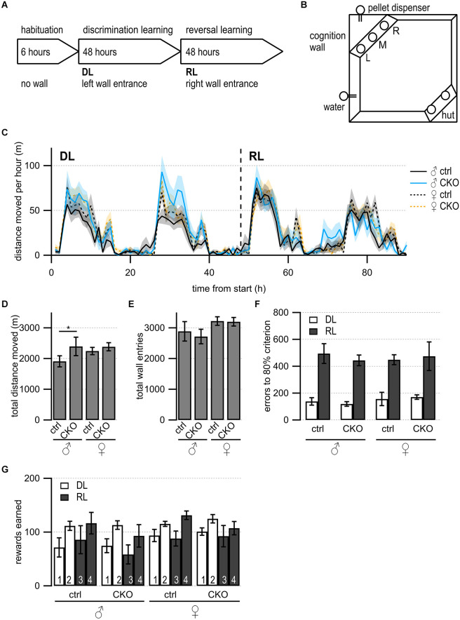
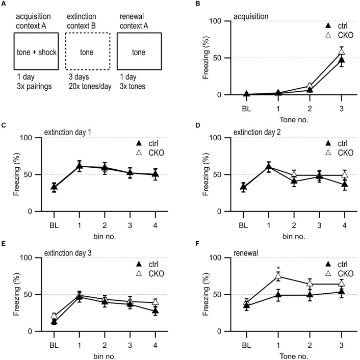
Introduction: Altered signaling or function of acetylcholine (ACh) has been reported in various neurological diseases, including Alzheimer's disease, Tourette syndrome, epilepsy among others. Many neurons that release ACh also co-transmit the neurotransmitter gamma-aminobutyrate (GABA) at synapses in the hippocampus, striatum, substantia nigra, and medial prefrontal cortex (mPFC). Although ACh transmission is crucial for higher brain functions such as learning and memory, the role of co-transmitted GABA from ACh neurons in brain function remains unknown. Thus, the overarching goal of this study was to investigate how a systemic loss of GABA co-transmission from ACh neurons affected the behavioral performance of mice. Methods: To do this, we used a conditional knock-out mouse of the vesicular GABA transporter (vGAT) crossed with the ChAT-Cre driver line to selectively ablate GABA co-transmission at ACh synapses. In a comprehensive series of standardized behavioral assays, we compared Cre-negative control mice with Cre-positive vGAT knock-out mice of both sexes. Results: Loss of GABA co-transmission from ACh neurons did not disrupt the animal's sociability, motor skills or sensation. However, in the absence of GABA co-transmission, we found significant alterations in social, spatial and fear memory as well as a reduced reliance on striatum-dependent response strategies in a T-maze. In addition, male conditional knockout (CKO) mice showed increased locomotion. Discussion: Taken together, the loss of GABA co-transmission leads to deficits in higher brain functions and behaviors. Therefore, we propose that ACh/GABA co-transmission modulates neural circuitry involved in the affected behaviors.

Keywords: GABA; acetylcholine; animal behavior; co-transmission; transgenic mice.


Figures

Figure 1
Figure 1
Loss of GABA co-transmission from ACh neurons results in loss of social novelty preference independent of sex as well as increased chamber entries in males. Loss of GABA co-transmission leads to a loss of social novelty preference in mice as well as increased chamber entries in males but does not affect sociability. (A) Schematic showing setup of sociability and social novelty preference test. Mice habituated for 10 min in the cage with both side chambers empty. Before the sociability test, an unfamiliar sex-matched mouse (stranger 1) was introduced into the left chamber. After 10 min of the sociability test, before the start of the social novelty preference test, an unfamiliar sex-matched mouse (stranger 2) was introduced into the right chamber. Time spent in outer chambers and chamber entries were measured. (B,C) Sociability test results. Time spent in (B) and entries (C) into side chambers shown for male, female control (ctrl), and CKO mice. (D,E) Social novelty test results. Time spent in (D) and entries (E) into side chambers shown for male, female control (ctrl), and CKO mice. Values represent mean ± SEM. Individual data points are depicted as open circles. Repeated measures ANOVA followed by Fisher’s protected least-significant difference tests (*p < 0.05, **p < 0.01, ***p < 0.001, ****p < 0.0001). See Supplementary Table 2 for data.
Figure 2
Figure 2
Loss of GABA co-transmission from ACh neurons results in spatial memory deficits. Loss of GABA co-transmission leads to spatial memory impairments. (A) Timeline of Morris water maze experiment. Visible platform test, acquisition test using hidden platform followed by one probe trial without platform, reversal learning test with hidden platform in the opposite quadrant, and one probe trial without platform. (B) Water maze setup for acquisition test. Visible/hidden platform placed at the circle. Start points chosen in arbitrarily random order. Theoretical escape pathway to hidden platform indicated by arrows. (C) Water maze setup for reversal test with hidden platform placed in opposite quadrant. (D,E) Latency to reach the hidden platform during acquisition learning for males (D) and females (E). (F) Probe trial results after acquisition learning phase in absence of the platform. Counts of target and opposite quadrant crosses. (G,H) Latency to reach the hidden platform during reversal learning for males (D) and females (E). (I) Probe trial results after reversal learning phase in absence of the platform. Counts of swimming path crosses over the platform area in target quadrant and corresponding area in the opposite quadrant. Values represent mean ± SEM. Individual data points are depicted as open circles. Repeated measures ANOVA followed by Fisher’s protected least-significant difference (*p < 0.05, **p < 0.01). See Supplementary Table 3 for data.
Figure 3
Figure 3
Loss of GABA co-transmission from ACh neurons leads to changes in usage of competing learning strategies. (A) Timeline of T maze experiment. Habituation phase for 2 days, training phase for 7 days followed by one probe trial day. Repetition of the training phase for another 7 days followed by one probe trial day. (B) T-maze setup. Animals start in the south arm during habituation and training but from the north arm during probe trials. Visible fabric cues attached to the curtained walls outside of the maze. Food bowls (circle) were placed at ends of both goal arms. Start points for training indicated by continuous arrow and for probe trials by dashed arrow. (C) Training success indicated as correct entries into goal arms for every training day. (D,E) Results for Probe trial day 1 (D) and Probe trial day 2 (E) indicating the relative number of response (R) and place (P) learners. (F) Animal ratios were sorted by learning strategy and strategy transitions from probe trial 1 to probe trial 2: place transitioner (R→P, green), consistent place (orange), consistent response (purple), response transitioner (P→R, turquoise). Values represent mean ± SEM. Repeated Measures Proportional Odds Logistic Regression model (C) and Fisher’s exact test (F) (*p < 0.05). See Supplementary Table 4 for data.
Figure 4
Figure 4
Loss of GABA co-transmission from ACh neurons results in increased locomotion in males but no impairments in discrimination or reversal learning. (A) Timeline of Cognition Wall experiment. Habituation phase for 6 h without Cognition Wall. Introduction of the Cognition Wall ~30 min before the start of discrimination learning phase (DL) for 2 days followed by reversal learning phase (RL) for another 2 days. During DL, the animal trained to enter left wall entrance for food reward. During RL, the animal trained to enter right wall entrance for food reward. (B) Cognition Wall setup. Hut for shelter in bottom right, water bottle in bottom left, Cognition Wall in top left covering the food dispenser. Left (L), middle (M), right (R) entrance of Cognition Wall. (C) The total distance moved during experiment per 1 h bin. (D) The total distance moved during experiment. (E) The total wall entries during experiment. (F) The number of error entries before reaching 80% learning criterion during DL and RL. (G) The number of reward pellets earned per day during DL and RL. Values represent mean ± SEM. Two-way ANOVA followed by Fisher’s protected least-significant difference (*p < 0.05). See Supplementary Table 5 for data.
Figure 5
Figure 5
Loss of GABA co-transmission from ACh neurons leads to transient increased freezing during contextual fear renewal. (A) Timeline of fear conditioning experiment. Acquisition of fear memory on day 1 by 3× pairing of 75 dB 2,800 Hz pure tone with 2 s 0.5 mV foot shock in context A. During 3 days of extinction, animal was presented with 20 tones/day in context B. During the renewal of the fear memory, animal was presented with three tones in context A. (B) Percent of freezing during acquisition phase at baseline (BL) and per tone/foot shock pairing. (C–E) Percent of freezing during extinction phase at baseline (BL) and per five tone bin on day 1 (C), day 2 (D), and day 3 (E). (F) Percent of freezing during renewal phase at baseline (BL) and per tone presentation. Values represent mean ± SEM. Two-way ANOVA followed by Fisher’s protected least-significant difference test (*p < 0.05). See Supplementary Table 6 for data.
Figure 6
Figure 6
Summary/simplified model. Simplified model of how ACh/GABA co-transmission is embedded in the circuitry of cortical and subcortical structures. ACh/GABA projections from the basal forebrain (MS/DBB and NB) form synapses in the hippocampus and mPFC, respectively. The hippocampal outputs vary based on dorsal or ventral location of the neurons to mPFC or EC, among others. Basal ganglia structures such as CPu or GPe contain either local ACh/GABA interneurons, receive inputs from LDT/PPN, or project to the mPFC. The mPFC contains local ACh/GABA interneurons and receives noradrenergic (NA) inputs from the locus coeruleus (LC). SNc DA neurons receive ACh/GABA projections from LDT/PPN.

Similar articles

Cited by

References

    1. Adkins J. M., Lynch J., Gray M., 3rd, Jasnow A. M. (2021). Presynaptic GABAB receptor inhibition sex dependently enhances fear extinction attenuates fear renewal. Psychopharmacology (Berl) 238, 2059–2071. 10.1007/s00213-021-05831-w - DOI - PMC - PubMed
    1. Albrecht A., Müller I., Weiglein A., Pollali E., Çalşkan G., Stork O. (2022). Choosing memory retrieval strategies: a critical role for inhibition in the dentate gyrus. Neurobiol. Stress 20:100474. 10.1016/j.ynstr.2022.100474 - DOI - PMC - PubMed
    1. Alexander G. M., Brown L. Y., Farris S., Lustberg D., Pantazis C., Gloss B., et al. . (2018). CA2 neuronal activity controls hippocampal low gamma and ripple oscillations. eLife 7:e38052. 10.7554/eLife.38052 - DOI - PMC - PubMed
    1. Alexander G. M., Farris S., Pirone J. R., Zheng C., Colgin L. L., Dudek S. M., et al. . (2016). Social novel contexts modify hippocampal CA2 representations of space. Nat. Commun. 7:10300. 10.1038/ncomms10300 - DOI - PMC - PubMed
    1. Amitai N., Young J. W., Higa K., Sharp R. F., Geyer M. A., Powell S. B., et al. . (2014). Isolation rearing effects on probabilistic learning cognitive flexibility in rats. Cogn. Affect. Behav. Neurosci. 14, 388–406. 10.3758/s13415-013-0204-4 - DOI - PMC - PubMed

LinkOut - more resources