

Advances in small molecule two-photon fluorescent trackers for lipid droplets in live sample imaging
- PMID: 36505737
- PMCID: PMC9733596
- DOI: 10.3389/fchem.2022.1072143
Advances in small molecule two-photon fluorescent trackers for lipid droplets in live sample imaging
Abstract
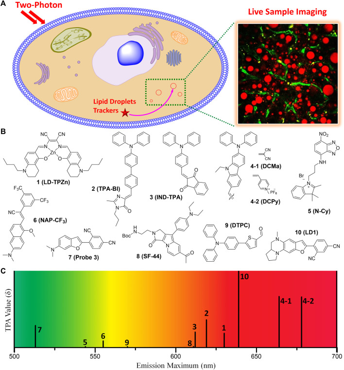
Two-photon fluorescent trackers for monitoring of lipid droplets (LDs) would be highly effective for illustrating the critical roles of LDs in live cells or tissues. Although a number of one-photon fluorescent trackers for labeling LDs have been developed, their usability remains constrained in live sample imaging due to photo damage, shallow imaging depth, and auto-fluorescence. Recently, some two-photon fluorescent trackers for LDs have been developed to overcome these limitations. In this mini-review article, the advances in two-photon fluorescent trackers for monitoring of LDs are summarized. We summarize the chemical structures, two-photon properties, live sample imaging, and biomedical applications of the most recent representative two-photon fluorescent trackers for LDs. Additionally, the current challenges and future research trends for the two-photon fluorescent trackers of LDs are discussed.
Keywords: lipid droplet; lipid metabolism; live sample imaging; organelle tracker; two-photon microscopy.
Copyright © 2022 Lee, Kim, Lee and Kim.
Conflict of interest statement
The authors declare that the research was conducted in the absence of any commercial or financial relationships that could be construed as a potential conflict of interest.
Figures
Similar articles
-
Recent advances in fluorescent probes for lipid droplets.Chem Commun (Camb). 2022 Feb 1;58(10):1495-1509. doi: 10.1039/d1cc05717k. Chem Commun (Camb). 2022. PMID: 35019910 Review.
-
Interface-Targeting Strategy Enables Two-Photon Fluorescent Lipid Droplet Probes for High-Fidelity Imaging of Turbid Tissues and Detecting Fatty Liver.ACS Appl Mater Interfaces. 2018 Apr 4;10(13):10706-10717. doi: 10.1021/acsami.8b00278. Epub 2018 Mar 22. ACS Appl Mater Interfaces. 2018. PMID: 29521495
-
Recent Advances in Fluorescent Probes for Lipid Droplets.Materials (Basel). 2018 Sep 18;11(9):1768. doi: 10.3390/ma11091768. Materials (Basel). 2018. PMID: 30231571 Free PMC article. Review.
-
Monitoring Lipid Droplet Dynamics in Living Cells by Using Fluorescent Probes.Biochemistry. 2019 Feb 12;58(6):499-503. doi: 10.1021/acs.biochem.8b01071. Epub 2019 Jan 11. Biochemistry. 2019. PMID: 30628446
-
Polarity-Driven Two-Photon Fluorescent Probe for Monitoring the Perturbation in Lipid Droplet Levels during Mitochondrial Dysfunction and Acute Pancreatitis.ACS Sens. 2023 Oct 27;8(10):3793-3803. doi: 10.1021/acssensors.3c01245. Epub 2023 Oct 10. ACS Sens. 2023. PMID: 37815484
Cited by
-
The application of infrared thermography technology in flap: A perspective from bibliometric and visual analysis.Int Wound J. 2023 Dec;20(10):4308-4327. doi: 10.1111/iwj.14333. Epub 2023 Aug 8. Int Wound J. 2023. PMID: 37551726 Free PMC article.
References
-
- Abramczyk H., Surmacki J., Kopec M., Olejnik A. K., Lubecka-Pietruszewska K., Fabianowska-Majewska K. (2015). The role of lipid droplets and adipocytes in cancer. Raman imaging of cell cultures: MCF10A, MCF7, and MDA-MB-231 compared to adipocytes in cancerous human breast tissue. Analyst 140 (7), 2224–2235. 10.1039/c4an01875c - DOI - PubMed
Publication types
LinkOut - more resources
Full Text Sources