Evaluation of diagnostic efficacy of multimode ultrasound in BI-RADS 4 breast neoplasms and establishment of a predictive model
- PMID: 36505867
- PMCID: PMC9730703
- DOI: 10.3389/fonc.2022.1053280
Evaluation of diagnostic efficacy of multimode ultrasound in BI-RADS 4 breast neoplasms and establishment of a predictive model
Abstract
Objectives: To explore the diagnostic efficacy of ultrasound (US), two-dimensional and three-dimensional shear-wave elastography (2D-SWE and 3D-SWE), and contrast-enhanced ultrasound (CEUS) in breast neoplasms in category 4 based on the Breast Imaging Reporting and Data System (BI-RADS) from the American College of Radiology (ACR) and to develop a risk-prediction nomogram based on the optimal combination to provide a reference for the clinical management of BI-RADS 4 breast neoplasms.
Methods: From September 2021 to April 2022, a total of 104 breast neoplasms categorized as BI-RADS 4 by US were included in this prospective study. There were 78 breast neoplasms randomly assigned to the training cohort; the area under the receiver-operating characteristic curve (AUC), 95% confidence interval (95% CI), sensitivity, specificity, positive predictive value (PPV), and negative predictive value (NPV) of 2D-SWE, 3D-SWE, CEUS, and their combination were analyzed and compared. The optimal combination was selected to develop a risk-prediction nomogram. The performance of the nomogram was assessed by a validation cohort of 26 neoplasms.
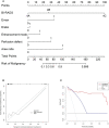
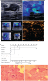
Results: Of the 78 neoplasms in the training cohort, 16 were malignant and 62 were benign. Among the 26 neoplasms in the validation cohort, 6 were malignant and 20 were benign. The AUC values of 2D-SWE, 3D-SWE, and CEUS were not significantly different. After a comparison of the different combinations, 2D-SWE+CEUS showed the optimal performance. Least absolute shrinkage and selection operator (LASSO) regression was used to filter the variables in this combination, and the variables included Emax, Eratio, enhancement mode, perfusion defect, and area ratio. Then, a risk-prediction nomogram with BI-RADS was built. The performance of the nomogram was better than that of the radiologists in the training cohort (AUC: 0.974 vs. 0.863). In the validation cohort, there was no significant difference in diagnostic accuracy between the nomogram and the experienced radiologists (AUC: 0.946 vs. 0.842).
Conclusions: US, 2D-SWE, 3D-SWE, CEUS, and their combination could improve the diagnostic efficiency of BI-RADS 4 breast neoplasms. The diagnostic efficacy of US+3D-SWE was not better than US+2D-SWE. US+2D-SWE+CEUS showed the optimal diagnostic performance. The nomogram based on US+2D-SWE+CEUS performs well.
Keywords: breast neoplasms; contrast media; elastography; nomograms; ultrasound.
Copyright © 2022 Chen, Lu, Li, Liao, Huang and Zhang.
Conflict of interest statement
The authors declare that the research was conducted in the absence of any commercial or financial relationships that could be construed as a potential conflict of interest.
Figures


Similar articles
-
The application of multimodal ultrasound examination in the differential diagnosis of benign and malignant breast lesions of BI-RADS category 4.Front Med (Lausanne). 2025 Jun 9;12:1596100. doi: 10.3389/fmed.2025.1596100. eCollection 2025. Front Med (Lausanne). 2025. PMID: 40552183 Free PMC article.
-
Predictive value of contrast-enhanced ultrasonography and ultrasound elastography for management of BI-RADS category 4 nonpalpable breast masses.Eur J Radiol. 2024 Apr;173:111391. doi: 10.1016/j.ejrad.2024.111391. Epub 2024 Feb 28. Eur J Radiol. 2024. PMID: 38422608
-
BI-RADS 4 breast lesions: could multi-mode ultrasound be helpful for their diagnosis?Gland Surg. 2019 Jun;8(3):258-270. doi: 10.21037/gs.2019.05.01. Gland Surg. 2019. PMID: 31328105 Free PMC article.
-
Diagnostic Performance of Contrast-Enhanced Ultrasound in the Evaluation of Small Renal Masses: A Systematic Review and Meta-Analysis.Diagnostics (Basel). 2022 Sep 25;12(10):2310. doi: 10.3390/diagnostics12102310. Diagnostics (Basel). 2022. PMID: 36291999 Free PMC article. Review.
-
A Review on Biological Effects of Ultrasounds: Key Messages for Clinicians.Diagnostics (Basel). 2023 Feb 23;13(5):855. doi: 10.3390/diagnostics13050855. Diagnostics (Basel). 2023. PMID: 36899998 Free PMC article. Review.
Cited by
-
Diagnostic value of contrast-enhanced ultrasound and shear-wave elastography for small breast nodules.PeerJ. 2024 Jul 3;12:e17677. doi: 10.7717/peerj.17677. eCollection 2024. PeerJ. 2024. PMID: 38974410 Free PMC article.
-
A comparative study on the features of breast sclerosing adenosis and invasive ductal carcinoma via ultrasound and establishment of a predictive nomogram.Front Oncol. 2023 Oct 23;13:1276524. doi: 10.3389/fonc.2023.1276524. eCollection 2023. Front Oncol. 2023. PMID: 37936612 Free PMC article.
-
Diagnostic performance of contrast-enhanced ultrasound combined with shear wave elastography in differentiating benign from malignant breast lesions: a systematic review and meta-analysis.Gland Surg. 2023 Nov 24;12(11):1610-1623. doi: 10.21037/gs-23-333. Epub 2023 Oct 20. Gland Surg. 2023. PMID: 38107493 Free PMC article.
-
Combined use of super-resolution ultrasound imaging and shear-wave elastography for differential diagnosis of breast masses.Front Oncol. 2024 Dec 20;14:1497140. doi: 10.3389/fonc.2024.1497140. eCollection 2024. Front Oncol. 2024. PMID: 39759128 Free PMC article.
References
LinkOut - more resources
Full Text Sources
Miscellaneous

