. 2022 Nov 23:12:1027914.
doi: 10.3389/fonc.2022.1027914. eCollection 2022.

Proteomic profiling of cell line-derived extracellular vesicles to identify candidate circulatory markers for detection of gallbladder cancer

Affiliations

Proteomic profiling of cell line-derived extracellular vesicles to identify candidate circulatory markers for detection of gallbladder cancer

Ratna Priya et al. Front Oncol. .

Abstract

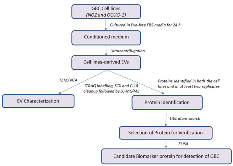
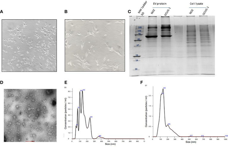
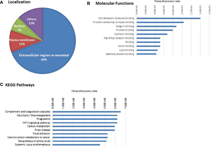
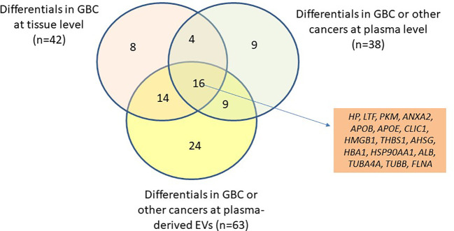
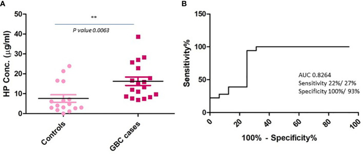
Gallbladder cancer (GBC) is the sixth most common gastrointestinal tract cancer with a very low overall survival and poor prognosis. Profiling of cancer-derived extracellular vesicles (EVs) is an emerging strategy for identification of candidate biomarkers for the detection and prognosis of the disease. The aim of the study was to analyse the protein content from GBC cell line- derived EVs with emphasis on proteins which could be used as candidate biomarkers for the detection of GBC. NOZ and OCUG-1 cell lines were cultured and EVs were isolated from conditioned media. LC-MS/MS analysis of total EV proteins led to the identification of a total of 268 proteins in both the cell lines. Of these, 110 proteins were identified with ≥2 unique peptides with ≥2 PSMs in at least two experimental and technical replicate runs. STRING (Search Tool for the Retrieval of Interacting Genes/Proteins) database was used to perform bioinformatics analysis of 110 proteins which showed 'cell adhesion molecule binding', 'integrin binding', 'cadherin binding' among the top molecular functions and 'focal adhesion' to be among the top pathways associated with the EV proteins. A total of 42 proteins including haptoglobin (HP), pyruvate kinase (PKM), annexin A2 (ANXA2), thrombospondin 1 (THBS1), were reported to be differentially abundant in GBC tissue. Of these, 16 proteins were reported to be differentially abundant in plasma and plasma-derived EVs. We infer these proteins to be highly important to be considered as potential circulatory biomarkers for the detection of GBC. To check the validity of this hypothesis, one of the proteins, haptoglobin (HP) as a representative case, was analysed in plasma by quantitative Enzyme- linked immunosorbent assay (ELISA) and we observed its increased levels in GBC in comparison to controls (p value= 0.0063). Receiver operating characteristic (ROC) curve analysis for GBC vs controls showed an Area under the ROC Curve (AUC) of 0.8264 for HP with 22% sensitivity against 100% specificity. We propose that HP along with other candidate proteins may be further explored for their clinical application.

Keywords: NOZ; OCUG-1; extracellular vesicles; gallbladder cancer (GBC); haptoglobin; proteomics.


Conflict of interest statement

The authors declare that the research was conducted in the absence of any commercial or financial relationships that could be construed as a potential conflict of interest.

Figures

Figure 1
Figure 1
Workflow of the study. GBC, Gallbladder carcinoma; EV, Extracellular Vesicles; iTRAQ, Isobaric tags for relative and absolute quantitation; ELISA, Enzyme-linked immunosorbent assay.
Figure 2
Figure 2
Characterization of GBC cell line-derived EVs. Representative image of GBC cell line NOZ (A) and OCUG-1 (B). SDS-PAGE profile of GBC cell line-derived EV proteins and cell lysate (C). Transmission electron micrographs of cell line- derived EVs. EVs from cell lines were isolated by ultracentrifugation method were resuspended in PBS and loaded on 2% carbon coated grids. Negative staining was performed using 2% phosphotungstic acid (PTA). Images of EVs (40-100 nm) were acquired using 200KV, TECNAI G20 HR-TEM, Thermo Scientific at 1,00,000× magnification, scale bar- 100 nm (D) Size and particle distribution plots of EVs from GBC cell line-derived EVs using nanoparticle tracking system showed peaks at 140 nm and 139 nm for NOZ (E) and OCUG-1 (F) respectively suggesting enrichment of exosomes in the EV fraction.
Figure 3
Figure 3
Gene ontology analysis of 110 cell line-derived EV proteins. (A) Localization (B) Molecular Function (C) KEGG pathways. The proteins associated with the top 10 molecular functions and pathways are provided in Supplementary Tables S5A and S5B .
Figure 4
Figure 4
Venn diagram showing the differential level of 110 proteins in tissue (GBC) and in plasma/serum or plasma/serum-derived EVs (GBC or other cancers). The differential level of 16 proteins were already reported in tissue, plasma/serum and plasma/serum-derived EVs and are candidate proteins to be explored as circulatory markers for GBC. HP, Haptoglobin; LTF, lactotransferrin isoform 1; PKM, pyruvate kinase isoform X1; ANXA2, annexin A2; APOB, apolipoprotein B-100; APOE, apolipoprotein E isoform a; CLIC1, chloride intracellular channel protein 1; HMGB1, high mobility group protein B1 isoform X1; THBS1, thrombospondin-1; AHSG, alpha-2-HS-glycoprotein isoform 2; HBA1, hemoglobin subunit alpha; HSP90AA1, heat shock protein HSP 90-alpha isoform 1; ALB, serum albumin; TUBA4A, tubulin alpha-4A chain isoform 1; TUBB, tubulin beta chain isoform b; FLNA, Filamin-A isoform 2.
Figure 5
Figure 5
Plasma level of HP in GBC cases and controls using quantitative ELISA. (A) Scatter plot showing concentration in plasma samples. Controls include healthy individuals and GSD cases. A significant increase in the levels of HP was observed in GBC cases (B) ROC curve representing sensitivity and specificity for HP. ** p value is less than or equal to 0.01.

Similar articles

Cited by

References

    1. World Health Organization (WHO) . Global health estimates 2020: Deaths by cause, age, sex, by country and by region, 2000-2019 (2020). WHO; (Accessed December 11, 2020).
    1. Montalvo-Jave EE, Rahnemai-Azar AA, Papaconstantinou D, Deloiza ME, Tsilimigras DI, Moris D, et al. . Molecular pathways and potential biomarkers in gallbladder cancer: A comprehensive review. Surg Oncol (2019) 31:83–9. doi: 10.1016/j.suronc.2019.09.006 - DOI - PubMed
    1. Andrén-Sandberg A. Diagnosis and management of gallbladder cancer. N Am J Med Sci (2012) 4(7):293–9. doi: 10.4103/1947-2714.98586 - DOI - PMC - PubMed
    1. Singh TD, Barbhuiya MA, Poojary S, Shrivastav BR, Tiwari PK. The liver function test enzymes and glucose level are positively correlated in gallbladder cancer: a cancer registry data analysis from north central India. Indian J Cancer (2012) 49(1):125–36. doi: 10.4103/0019-509X.98938 - DOI - PubMed
    1. Yáñez-Mó M, Siljander PR, Andreu Z, Zavec AB, Borràs FE, Buzas EI, et al. . Biological properties of extracellular vesicles and their physiological functions. J Extracell Vesicles (2015) 4:27066. doi: 10.3402/jev.v4.27066 - DOI - PMC - PubMed