

Predictive, preventive, and personalized management of retinal fluid via computer-aided detection app for optical coherence tomography scans
- PMID: 36505893
- PMCID: PMC9727042
- DOI: 10.1007/s13167-022-00301-5
Predictive, preventive, and personalized management of retinal fluid via computer-aided detection app for optical coherence tomography scans
Abstract
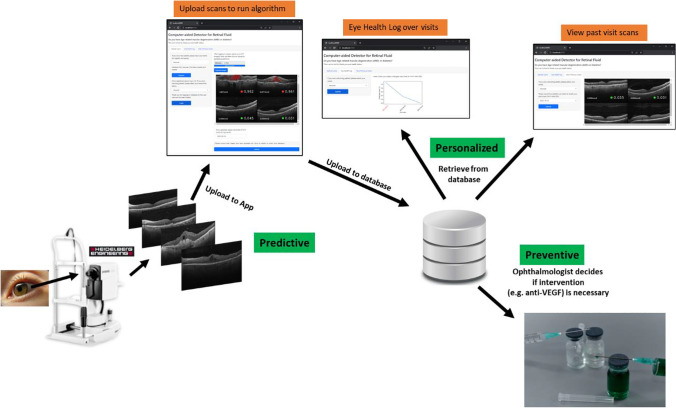
Aims: Computer-aided detection systems for retinal fluid could be beneficial for disease monitoring and management by chronic age-related macular degeneration (AMD) and diabetic retinopathy (DR) patients, to assist in disease prevention via early detection before the disease progresses to a "wet AMD" pathology or diabetic macular edema (DME), requiring treatment. We propose a proof-of-concept AI-based app to help predict fluid via a "fluid score", prevent fluid progression, and provide personalized, serial monitoring, in the context of predictive, preventive, and personalized medicine (PPPM) for patients at risk of retinal fluid complications.
Methods: The app comprises a convolutional neural network-Vision Transformer (CNN-ViT)-based segmentation deep learning (DL) network, trained on a small dataset of 100 training images (augmented to 992 images) from the Singapore Epidemiology of Eye Diseases (SEED) study, together with a CNN-based classification network trained on 8497 images, that can detect fluid vs. non-fluid optical coherence tomography (OCT) scans. Both networks are validated on external datasets.
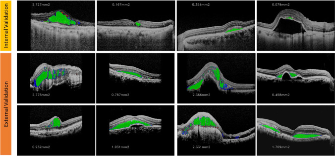
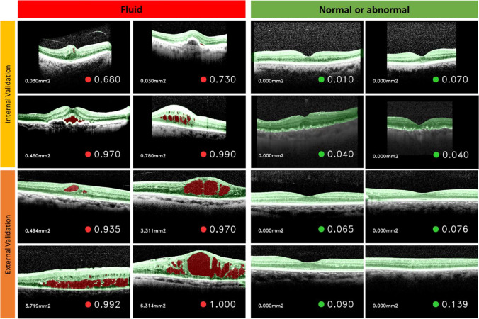
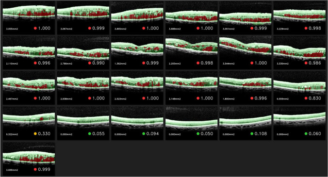
Results: Internal testing for our segmentation network produced an IoU score of 83.0% (95% CI = 76.7-89.3%) and a DICE score of 90.4% (86.3-94.4%); for external testing, we obtained an IoU score of 66.7% (63.5-70.0%) and a DICE score of 78.7% (76.0-81.4%). Internal testing of our classification network produced an area under the receiver operating characteristics curve (AUC) of 99.18%, and a Youden index threshold of 0.3806; for external testing, we obtained an AUC of 94.55%, and an accuracy of 94.98% and an F1 score of 85.73% with Youden index.
Conclusion: We have developed an AI-based app with an alternative transformer-based segmentation algorithm that could potentially be applied in the clinic with a PPPM approach for serial monitoring, and could allow for the generation of retrospective data to research into the varied use of treatments for AMD and DR. The modular system of our app can be scaled to add more iterative features based on user feedback for more efficient monitoring. Further study and scaling up of the algorithm dataset could potentially boost its usability in a real-world clinical setting.
Supplementary information: The online version contains supplementary material available at 10.1007/s13167-022-00301-5.
Keywords: Age-related macular degeneration; Anti-vascular endothelial growth factor; Computer-aided detection; Deep learning; Diabetic macular edema; Diabetic retinopathy; Ophthalmology; Optical coherence tomography; Predictive, preventive, and personalized OCT; Retinal fluid.
© The Author(s), under exclusive licence to European Association for Predictive, Preventive and Personalised Medicine (EPMA) 2022, Springer Nature or its licensor (e.g. a society or other partner) holds exclusive rights to this article under a publishing agreement with the author(s) or other rightsholder(s); author self-archiving of the accepted manuscript version of this article is solely governed by the terms of such publishing agreement and applicable law.
Conflict of interest statement
Competing interestsT.H.R. was a former scientific adviser and owns stock of Medi Whale. All other authors declare no competing interests.
Figures


Similar articles
-
Automated Segmentation of Retinal Fluid Volumes From Structural and Angiographic Optical Coherence Tomography Using Deep Learning.Transl Vis Sci Technol. 2020 Oct 8;9(2):54. doi: 10.1167/tvst.9.2.54. eCollection 2020 Oct. Transl Vis Sci Technol. 2020. PMID: 33110708 Free PMC article.
-
OCT-based deep learning algorithm for the evaluation of treatment indication with anti-vascular endothelial growth factor medications.Graefes Arch Clin Exp Ophthalmol. 2018 Jan;256(1):91-98. doi: 10.1007/s00417-017-3839-y. Epub 2017 Nov 10. Graefes Arch Clin Exp Ophthalmol. 2018. PMID: 29127485
-
Evaluation of an Artificial Intelligence-Based Detector of Sub- and Intraretinal Fluid on a Large Set of Optical Coherence Tomography Volumes in Age-Related Macular Degeneration and Diabetic Macular Edema.Ophthalmologica. 2022;245(6):516-527. doi: 10.1159/000527345. Epub 2022 Oct 10. Ophthalmologica. 2022. PMID: 36215958
-
Advancing Diabetic Retinopathy Diagnosis: Leveraging Optical Coherence Tomography Imaging with Convolutional Neural Networks.Rom J Ophthalmol. 2023 Oct-Dec;67(4):398-402. doi: 10.22336/rjo.2023.63. Rom J Ophthalmol. 2023. PMID: 38239418 Free PMC article. Review.
-
Recent Advanced Deep Learning Architectures for Retinal Fluid Segmentation on Optical Coherence Tomography Images.Sensors (Basel). 2022 Apr 15;22(8):3055. doi: 10.3390/s22083055. Sensors (Basel). 2022. PMID: 35459040 Free PMC article. Review.
Cited by
-
Development of a deep learning model to distinguish the cause of optic disc atrophy using retinal fundus photography.Sci Rep. 2024 Mar 1;14(1):5079. doi: 10.1038/s41598-024-55054-0. Sci Rep. 2024. PMID: 38429319 Free PMC article.
-
Artificial Intelligence-Based Quantification of Central Macular Fluid Volume and VA Prediction for Diabetic Macular Edema Using OCT Images.Ophthalmol Ther. 2023 Oct;12(5):2441-2452. doi: 10.1007/s40123-023-00746-5. Epub 2023 Jun 15. Ophthalmol Ther. 2023. PMID: 37318706 Free PMC article.
-
Development and validation of a routine blood parameters-based model for screening the occurrence of retinal detachment in high myopia in the context of PPPM.EPMA J. 2023 Mar 15;14(2):219-233. doi: 10.1007/s13167-023-00319-3. eCollection 2023 Jun. EPMA J. 2023. PMID: 37275550 Free PMC article.
-
Deep learning prediction of steep and flat corneal curvature using fundus photography in post-COVID telemedicine era.Med Biol Eng Comput. 2024 Feb;62(2):449-463. doi: 10.1007/s11517-023-02952-6. Epub 2023 Oct 27. Med Biol Eng Comput. 2024. PMID: 37889431
-
Discriminative, generative artificial intelligence, and foundation models in retina imaging.Taiwan J Ophthalmol. 2024 Nov 28;14(4):473-485. doi: 10.4103/tjo.TJO-D-24-00064. eCollection 2024 Oct-Dec. Taiwan J Ophthalmol. 2024. PMID: 39803410 Free PMC article. Review.
References
-
- Pichi, F. and P. Neri, Complications in uveitis. p. X, 288 p. 89 illus., 73 illus. in color. online resource.
LinkOut - more resources
Full Text Sources
