. 2022 Nov 25:13:989815.
doi: 10.3389/fendo.2022.989815. eCollection 2022.

Human stem cell derived beta-like cells engineered to present PD-L1 improve transplant survival in NOD mice carrying human HLA class I

Affiliations

Human stem cell derived beta-like cells engineered to present PD-L1 improve transplant survival in NOD mice carrying human HLA class I

Jorge Santini-González et al. Front Endocrinol (Lausanne). .

Abstract

There is a critical need for therapeutic approaches that combine renewable sources of replacement beta cells with localized immunomodulation to counter recurrence of autoimmunity in type 1 diabetes (T1D). However, there are few examples of animal models to study such approaches that incorporate spontaneous autoimmunity directed against human beta cells rather than allogenic rejection. Here, we address this critical limitation by demonstrating rejection and survival of transplanted human stem cell-derived beta-like cells clusters (sBCs) in a fully immune competent mouse model with matching human HLA class I and spontaneous diabetes development. We engineered localized immune tolerance toward transplanted sBCs via inducible cell surface overexpression of PD-L1 (iP-sBCs) with and without deletion of all HLA class I surface molecules via beta-2 microglobulin knockout (iP-BKO sBCs). NOD.HLA-A2.1 mice, which lack classical murine MHC I and instead express human HLA-A*02:01, underwent transplantation of 1,000 human HLA-A*02:01 sBCs under the kidney capsule and were separated into HLA-A2 positive iP-sBC and HLA-class I negative iP-BKO sBC groups, each with +/- doxycycline (DOX) induced PD-L1 expression. IVIS imaging showed significantly improved graft survival in mice transplanted with PD-L1 expressing iP-sBC at day 3 post transplantation compared to controls. However, luciferase signal dropped below in vivo detection limits by day 14 for all groups in this aggressive immune competent diabetes model. Nonetheless, histological examination revealed significant numbers of surviving insulin+/PD-L1+ sBCs cells for DOX-treated mice at day 16 post-transplant despite extensive infiltration with high numbers of CD3+ and CD45+ immune cells. These results show that T cells rapidly infiltrate and attack sBC grafts in this model but that significant numbers of PD-L1 expressing sBCs manage to survive in this harsh immunological environment. This investigation represents one of the first in vivo studies recapitulating key aspects of human autoimmune diabetes to test immune tolerance approaches with renewable sources of beta cells.

Keywords: HLA Class I; PD-L1; beta cell replacement; humanized mouse; stem cell-derived beta cell; tolerance; type 1 diabetes.


Conflict of interest statement

HR is or has been a consultant and islets SAB member to Sigilon therapeutics, a SAB member at Prellis Biologics and consultant to Eli Lilly and Minutia. The remaining authors declare that the research was conducted in the absence of any commercial or financial relationships that could be construed as a potential conflict of interest.

Figures
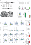
Figure 1
Figure 1
Quantification of HLA class I and PD-L1 expression on stem cell derived beta cells containing inducible PD-L1 expression with or without B2M knock out. (A) Schematic of the PD-L1 inducible system integrated into the AAVS1 locus of human pluripotent stem cells that contain a GFP fluorescence reporter under the control of the endogenous insulin promotor (INS-GFP). Constitutive expression of a puromycin resistance, a luciferase gene and a neomycin resistance gene is driven by the endogenous AAVS1 gene via T2A peptide cleavage sites on either allele, respectively. The transgene cassette also contains a strong CAG promoter driving constitutive expression of the M2rtTA that facilitates DOX inducible expression of PD-L1 under the control of the TRE3G promoter, bicistronicly. (B) Schematic of the CRISPR/Cas9 knock-out (KO) of B2M gene. Two gRNAs directed towards exon 1 and exon 2 of the B2M gene were used resulting in B2M gene KO and inhibition of HLA class I surface expression. (C) Schematic of the 3D suspension based directed differentiation protocol for the generation of stem cell derived beta cells (sBC) from human pluripotent stem cells (hPSC). (D) Bright field and fluorescence images of differentiated sBCs containing an endogenous insulin promoter driven GFP (INS-GFP) reporter. Scale bar represents 200 µm. (E) Representative flow cytometry analysis (left) and associated quantification (right) for beta cell markers cPEP and NKX6.1 of differentiated sBCs. Single positive cPEP and double positive cPEP and NKX6.1 cells are quantified. n = 4 independent experiments employing both iP and iP-BKO cells. (F) Representative HLA-ABC and INS-GFP flow cytometry analysis and associated quantification of differentiated iP sBC- (top row) and iP-BKO sBC- clusters (bottom row) untreated or treated with DOX (2 ug/ml), IFNg (100 ng/ml) or both for 48 hr. Bar plots represent HLA-ABC MFI quantification of INS-GFP+ cells from n = 3 independent differentiation experiments. One-Way ANOVA with multiple comparisons was used to determine significant changes between the groups (n = 3 for iP-BKO). (G) Representative PD-L1 and INS-GFP flow cytometry analysis and associated quantification of differentiated iP sBC- (top row) and iP-BKO sBC- clusters (bottom row) untreated or treated with DOX (2 ug/ml), IFNg (100 ng/ml) or both for 48 hr. Bar plots represent PD-L1 MFI quantification of INS-GFP+ cells of n = 3 independent differentiation experiments. One-Way ANOVA with multiple comparisons was used to determine significant changes between the groups (n=3 for iP-BKO). All error bars represent mean ± s.e.m. * p < 0.05 and ** p < 0.01. ns, Not significant.
Figure 2
Figure 2
Timeline of the study. HLA-A2 and BKO sBC clusters were generated with a Tet-On PD-L1 inducible system (iP) and constitutive luciferase expression. PD-L1 is expressed by the sBC clusters in presence of doxycyline (DOX). Four days prior to transplantation, mice were put on a special diet with doxycycline (+) or maintained on standard diet (-). About 1,000 sBC clusters were transplanted to the left kidney capsule of each mouse. Survival of the transplanted sBCs was monitored by transdermal luminescence detection with IVIS. The sBC graft-bearing kidneys were recovered after completion of the 2-week study period for histology. Created with Biorender.com.
Figure 3
Figure 3
Longitudinal monitoring of the sBC graft by transdermal bioluminescence detection via IVIS. (A, B) Representative images of bioluminescence expression by (A) iP-sBC or (B) iP-BKO sBC graft after transplant on day 1, 3, 7 or 9, and 14. (C, D) Quantification of the total flux measured from mice of both sex in the (C) iP-sBC and (D) iP-BKO sBC experiments. Each sBC group is subdivided by +/- DOX treatment as follows: iP (n=7), iP + DOX (n=8), iP-BKO (n=6) and iP-BKO + DOX (n=7). Two-Way ANOVA, *p < 0.05, **p-value < 0.01 mean ± s.e.m. (E) Kaplan-Meier curve for percentage of mice with detectable bioluminescence signal for all groups.
Figure 4
Figure 4
Insulin and PD-L1 expression by sBCs 2-weeks after transplantation. (A) Representative maximum intensity projection (MIP) image of whole graft-bearing kidney tissue section from the iP-sBC +DOX group immunostained for insulin, PD-L1 and CD3. (B, C) Expression and quantification of insulin and PD-L1 by iP and iP + DOX sBCs in inner graft zone. (D, E) Representative images and quantification of CD3+ cells in the outer graft zone of iP and iP + DOX sBCs. (F, G) Expression and quantification of insulin and PD-L1 by iP-BKO and iP-BKO + DOX sBCs in inner graft zone. (H, I) Representative images and quantification of CD3+ cells in the outer graft zone of iP-BKO and iP-BKO +DOX sBCs. The inner and outer graft zone in (A) are indicated with white dashed line and the regions enclosed in yellow dashed line depict location in the graft-bearing kidney from which the high magnification images shown of insulin and PD-L1 (B) or CD3 (D) were taken. n = 8 to 12 immunostained 10 μm thin sections of 2 to 4 recovered graft-bearing kidneys per condition, from which graft was successfully located. Two-tailed Student’s t-test, *p = 0.0394, **p = 0.0023, ***p = 0.0008, ****p < 0.0001, mean ± s.e.m. ns, Not significant.

Similar articles

Cited by

References

    1. Campbell-Thompson M, Fu A, Kaddis JS, Wasserfall C, Schatz DA, Pugliese A, et al. . Insulitis and beta-cell mass in the natural history of type 1 diabetes. Diabetes (2016) 65(3):719–31. doi: 10.2337/db15-0779 - DOI - PMC - PubMed
    1. Martin-Pagola A, Sisino G, Allende G, Dominguez-Bendala J, Gianani R, Reijonen H, et al. . Insulin protein and proliferation in ductal cells in the transplanted pancreas of patients with type 1 diabetes and recurrence of autoimmunity. Diabetologia (2008) 51(10):1803–13. doi: 10.1007/s00125-008-1105-x - DOI - PMC - PubMed
    1. Ryan EA, Lakey JR, Paty BW, Imes S, Korbutt GS, Kneteman NM, et al. . Successful islet transplantation: continued insulin reserve provides long-term glycemic control. Diabetes (2002) 51(7):2148–57. doi: 10.2337/diabetes.51.7.2148 - DOI - PubMed
    1. Ryan EA, Paty BW, Senior PA, Bigam D, Alfadhli E, Kneteman NM, et al. . Five-year follow-up after clinical islet transplantation. Diabetes (2005) 54(7):2060–9. doi: 10.2337/diabetes.54.7.2060 - DOI - PubMed
    1. Russ HA, Parent AV, Ringler JJ, Hennings TG, Nair GG, Shveygert M, et al. . Controlled induction of human pancreatic progenitors produces functional beta-like cells in vitro . EMBO J (2015) 34(13):1759–72. doi: 10.15252/embj.201591058 - DOI - PMC - PubMed